(1)
Department of Vascular Surgery, Evangelisches Krankenhaus Königin Elisabeth Herzberge, Berlin, Germany
In this chapter, by central veins we mean the axillary segment of the brachial, the axillary, the subclavian, the internal jugular, the brachiocephalic, the common femoral, the iliac veins, and the superior vena cava.
9.1 Pathophysiology
The causes of stenoses and occlusions differ according to their sites. The most frequent causes are:
Central venous catheters (subclavian, internal jugular, and femoral veins)
Unphysiological hemodynamic conditions near venous anastomoses of AV shunts (brachial, axillary, and subclavian veins)
Stents (cephalic and subclavian veins)
Chemotherapy
From March 2007 through June 2011 (51 months), 297 arm venograms were performed at our institution for malfunctioning AV accesses. In 129 patients we found stenoses and occlusions of central veins. Their topographic distribution is shown in Fig. 9.1. The subclavian vein is concerned strikingly often (right 22.2 %, left 11.4 %). All these patients had had subclavian catheters weeks to years previously. The central segments of the axillary veins on both sides showed 25 (13.5 %) stenoses or occlusions (Fig. 9.1, segment 3). Of these, 18 were caused by catheters and 7 by venous anastomoses of AV shunts. All 25 stenoses and occlusions (13.6 %) of the axillary segment of the brachial vein (Fig. 9.1, segment 1) can be traced back to venous anastomoses of AV shunts.
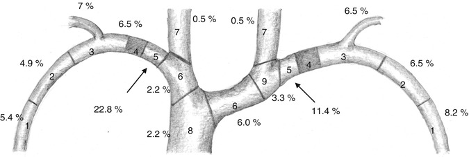
Fig. 9.1
Topographic distribution of stenoses and occlusions in 129 patients. 1 Axillary segment of the brachial vein, 2 peripheral segment of the axillary vein, 3 central segment of the axillary vein, 4 retroclavicular segment of the subclavian vein, 5 central segment of the subclavian vein, 6 brachiocephalic vein, 7 internal jugular vein, 8 superior vena cava, 9 confluence of the subclavian and left internal jugular veins
9.2 Clinical Signs and Investigations
Central venous stenoses or occlusions manifest themselves as:
Venous congestion with a patent AV access or
Thrombosis of the AV access
As the process of developing a stenosis or occlusion usually takes some time, subcutaneous venous collaterals, which do not have to be tightly filled, are frequently present. Diagnostics should include color duplex studies. Except for retroclavicular and retrosternal venous segments, reliable results may be obtained. With confirmed or suspected stenoses or occlusions of central veins, we also order venograms. Without a digital subtraction technique, however, contrast medium dilution may lead to an insufficient depiction of the large veins if contrast medium is injected into a (still) high flow AV access. Temporary flow reduction may be achieved by manual compression of the AV access distal to the injection site.
9.3 Therapy
The choice of treatment depends on:
Clinical impact of the stenosis or occlusion
Its location
The long-term prognosis of the AV access
Previous surgery in the region of the stenosis or occlusion
Prospects of endovascular therapy
Remaining options for the creation of future AV accessesRelated posts:

Stay updated, free articles. Join our Telegram channel

Full access? Get Clinical Tree


