Fig. 5.1
OR setting: S surgeon, A assistant, AN anesthesiologist, SN scrub nurse. Note the Y connector on the insufflator delivering CO2 to two separate lines
5.2 Access Port
The original technique used a 12-mm trocar that was put beside two Apple™ trocars (Apple Medical Corporation, Marlborough, MA, USA) through different fascial openings but through the same skin incision [1]. With three, very close to each other, fascial openings, there was a frequent loss of pneumoperitoneum, particularly when using large-bore instruments. This significantly reduced the quality of vision which, when too compromised, in fact made it impossible to continue with the intervention with the necessary safety.
The advent of trocars with multiple channels, such as SILS-Port™ (Covidien, Mansfield, MA, USA), TriPort™ (Olympus Medical Systems, Hamburg, Germany), and Octoport™ (Dalim, Seoul, Korea), has significantly reduced this inconvenience (Fig. 5.2). The main differences are the presence or absence of rigid cannulas, the number and size of accesses, and the adaptability to wall thickness.
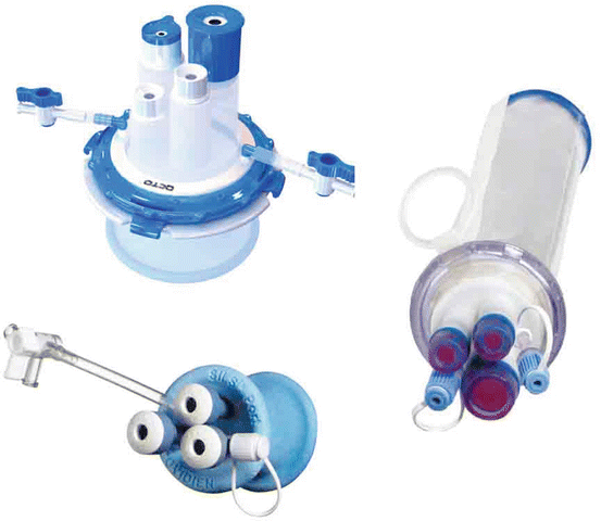
Fig. 5.2
Common single-incision access ports
In order to totally hide the scar after surgery, the umbilicus is completely everted as a glove finger, and an Ellis clamp is affixed at its base so as to demarcate the cut that will be about 12 mm. At this level, the fascia and the skin are very close to each other so it is easy to gain access to the peritoneal cavity with an open technique. When repositioned, the incision and its scar will not be visible (Fig. 5.3).
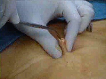
Fig. 5.3
Technique for umbilical incision. The umbilicus is everted as a glove finger. When repositioned, the incision will not be visible
5.3 Triangulation
The fundamental principle of single incision is instrument crossing in the navel where they enter the abdominal cavity. Thanks to the use of at least one articulated or curved instrument, paired with another instrument, either straight or articulated, it is possible to recreate a triangulation similar to that of conventional laparoscopy. As a consequence, the instrument held with the right hand is to be operated to the left on the screen and vice versa (Fig. 5.4).
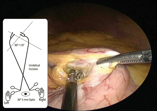
Fig. 5.4
The instruments cross each other at the umbilicus entrance point. With at least one curved or articulated instrument, it is possible to recreate an angle for traction. Note that the left hand is exerting traction, right side of the screen, while the right hand is dissecting the lesser curvature
5.4 Exposure
The use of a long shaft, 30-degree and 5-mm scope, is mandatory not only to reduce to a minimum the friction in the port but also to facilitate its movement during the course of the intervention and in order to reduce clashing of the instruments, particularly outside the abdomen.
The use of a camera with articulated head (EndoEYE™ – Olympus Medical Systems, Hamburg, Germany) furthermore reduces the need for these movements and offers optimal visual angles, thus avoiding conflict with surgical instruments: it therefore should be regarded as a very useful instrument for this type of approach [2] (Fig. 5.5).
Fig. 5.5
Surgeon position. The instruments are used in an upside position to avoid clashing and interference with the abdominal wall. Wrist and arm position are comfortable and without stress
With regard to bariatric surgery, the need to expose the gastroesophageal junction, to perform anastomosis and suture, has posed specific problems: some steps need to be modified so that, for example, the use of a liver retractor would not be necessary [2, 3].
Retraction of the left lobe of the liver can be achieved with a transfix stitch, applied on the right crus and suspended percutaneously.
5.5 Patient Selection
When new surgical techniques are adopted, selecting suitable patients is recommended. Although evaluating parameters like distance from the xifhoid may be simple, in practice, it might not be enough.
Relative contraindications, also in relation to the surgeon’s level of experience, such as a previous laparotomy or a previous laparoscopy, for which the navel was used as the access point, should be taken into account. In both cases, the difficulty of access or of positioning of the trocar in the peritoneal cavity should be verified. In fact, single-incision laparoscopy lacks the possibility of a provisional access for the camera at a point far from the navel or in any case of suspected adhesions, so that lysis can be performed to facilitate access.
In single-incision laparoscopy, the thickness of the abdominal wall is another feature to take into account as the port may not be sufficiently long to completely pass through it, and therefore, there might be a tendency to slide out, causing loss of the pneumoperitoneum and risk of damage when introducing the instrument.
A further consideration when selecting suitable patients is the position of the navel. The distance of the abdomen and hence the final position of the navel are difficult to determine in many cases, and it may be reasonably far from the initial position in the case of a large flaccid abdomen. In addition to that, the final distance at which the surgeon operates, such as the distance of the gastroesophageal junction from the navel, also depends on the patient’s height and physical build.
The possibility, even at present, of having dedicated and extra-long instruments available must be part of these preoperative evaluations, although intraoperative evaluation remains crucial.
5.6 Restrictive Procedures
Restrictive procedures based on reduction of food intake include the gastric band and the sleeve resection (Fig. 5.6).
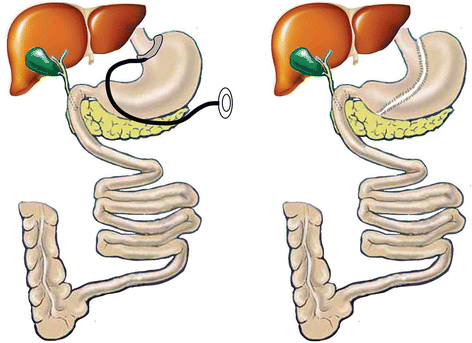
Fig. 5.6
Restrictive procedures. The band and the sleeve resection are based on creating an obstacle to passage of food or to reducing the capacity of the stomach
5.7 Sleeve Gastrectomy
Single-incision access and liver retraction are done as described above. The patient is placed in an anti-Trendelenburg position rotated to the right. The first step of the procedure is to dissect the angle of His to expose the left diaphragmatic pillar. Then we enter the lesser sac: LigaSure™ (Covidien, Mansfield, MA USA) 5 mm blunt tip 44 cm is used to open the gastroepiploic ligament enough to expose the posterior aspect of the angulus.
An orogastric calibration tube 40 Fr is inserted all the way to the pylorus. An Endo GIA™ (Covidien, Mansfield, MA USA) articulating 60 black reload is fired at a distance of about 6 cm from the pylorus toward the angulus. Suspension and correct traction of the stomach may require the use of transfix stitches passed through the stomach’s greater curvature and percutaneously in the left hypocondrium to suspend from the outside. The vertical transection of the stomach is continued with applications of Endo GIA™ Articulating 60 purple or tan reload along the orogastric tube. No reinforcement of the transection line is needed. Complete dissection of the gastroesophageal junction is of utmost importance. Then the section of the gastroepiploic ärtery branches is continued upward to the angle of His paying attention to stay close to the gastric wall and preserving the main artery branch. The patency and the tightness of the sleeve are tested with methylene blue. No drainage is left in place. The resected stomach is extracted, the pneumoperitoneum evacuated, and the port removed. The fascia is closed with an absorbable suture, and the navel is reinstated in its original position. We stress the pouch-first approach, which greatly facilitates the procedure particularly in single incision.
5.8 Gastric Band
Single-incision access and liver retraction is done as described above. The band-calibrated balloon-tipped orogastric tube is inserted into the stomach. Dissection of the angle of His is carried out as in the standard procedure using the SILS™ Clinch XL (Covidien, Mansfield, MA USA) in the left trocar and a regular hook in the right hand. A tunnel is created behind the stomach, as described for the pars flaccida technique. The dedicated band-articulating instrument is directed lateral to the equator of the calibration balloon and brought out at the angle of His. The band is closed around the stomach and the stoma calibrated using the inflated balloon (15 cm3). Three retention gastro-gastric nonabsorbable sutures are applied to prevent band and/or stomach slippage. At the end of the procedure, a supraumbilical subcutaneous tunnel is made, and the access port is fixed above the fascia. Careful reconstruction of the umbilicus will allow it to be placed back in its original position, thus achieving a completely invisible scar.
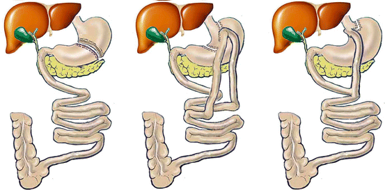
5.9 Mixed Procedures
5.9.1 Gastric Bypass
This very popular procedure consists of excluding from food transit part of the stomach, the duodenum, and variable part of the jejunum (Fig. 5.7). The gastric pouch size varies from 15 to 500 ml. In the Roux-en-Y gastric bypass, the pouch is very small and the bowel limbs relatively short. The weight loss is based on food intake reduction. In the biliopancreatic diversion, the pouch is very big with little or no food restriction, but most of the bowel is excluded from food transit. The weight loss is obtained by limiting food absorption. The minigastric bypass has an intermediate pouch and limb lengths. The food intake is partially limited as well as food absorption.
 Single-Access Laparoscopic Repair of Abdominal Wall Hernias
 Single-Incision Laparoscopic Appendectomy
 Single Access Laparoscopic Cholecystectomy
 Single-Access Laparoscopic Approach for Pancreatic Surgery
 Single-Access Laparoscopic Right Hemicolectomy
 Single-Access Laparoscopic Approach for Gastric Surgery (Hiatal Hernia Repair and Gastric Resections)


Related posts:
Single-Access Laparoscopic Repair of Abdominal Wall Hernias
Single-Incision Laparoscopic Appendectomy
Single Access Laparoscopic Cholecystectomy
Single-Access Laparoscopic Approach for Pancreatic Surgery
Single-Access Laparoscopic Right Hemicolectomy
Single-Access Laparoscopic Approach for Gastric Surgery (Hiatal Hernia Repair and Gastric Resections)
Stay updated, free articles. Join our Telegram channel
Full access? Get Clinical Tree
