Fig. 7.1
Intraoperative view of the multitrocar approach for a single-incision splenectomy
The technique used for splenic dissection is similar to that used in standard LS. After an explorative laparoscopy, the possible existence of accessory spleens is ruled out. A 5-mm curved grasper used for transanal endoscopic microsurgery (TEM) (Richard Wolf, Vernon Hills, IL, USA) is placed through the left port. The slightly curved end of this instrument fits into the flexible trocar or through a port of the multichannel device, and it is sufficiently curved to work intra-abdominally without causing instruments to clash. A 5-mm harmonic scalpel (Harmonic Ace, Ethicon Endo-Surgery, Cincinnati, OH, USA) is then introduced through the right port. Using this approach, it is possible to mobilize the splenic colon flexure and to reach the lower pole of the spleen. The next step is to gain access to the retrogastric pouch and to severe the short vessels at the upper pole of the spleen. With this view, and thanks to the flexible tip of the scope, it is possible, if desired, to ligate or clip the splenic artery. The instruments are then moved to the posterior face of the spleen, and the table is tilted to the left to take advantage of gravity and obtain exposure of the retrosplenic area. The posterior splenorenal attachments are freed.
Sometimes, especially if the umbilical approach is used and there are some difficulties with the more posterior and upper part of the upper splenic pole, a 3-mm instrument can be introduced through the left flank. This mini-instrument can be used to retract or section (hook) retroperitoneal adhesion.
Once the spleen is completely mobile, the flexible scope is retrieved, and the intra-abdominal visual control is changed to a 5-mm scope. If the multichannel has several large bore ports (Qadriport, Olympus), the 10-mm scope can be maintained. A probe inserted through the left 5-mm trocar raises the splenic hilum, providing sufficient space for the placement of the stapler. A stapler with a 6-cm white cartridge (Echelon, Ethicon Endo-Surgery) is inserted through a 12-mm trocar/port and advanced to the splenic fossa. After adjusting the jaws, the stapler is applied several times to sever the splenic hilum.
Once the spleen is completely free, a 15-mm endobag (Endo Catch II; Covidien, Mansfield, MA, USA) is inserted. The spleen is grasped with a 5-mm instrument and hung in the splenic fossa. The bag is deployed below the organ and the spleen is introduced. The bag is pulled to the umbilical incision and the spleen is retrieved (intact or morcellated), and the operating field is revised and complete hemostasis is achieved.
In the case of fenestration of a splenic cyst, the first step is to puncture and evacuate the cyst content. Then, with the help of the harmonic scalpel, we excise the maximum segment of the wall cyst, reaching the spleen parenchyma. Once hemostasis is completed, cyst wall fragments are extracted in an endobag.
The umbilicus is closed and carefully reconstructed, obtaining an optimal esthetic result.
7.3 Reduced Port Splenectomy
Single-port access splenectomy (SPAS) emphasizes the concept of surgery through one small transabdominal incision rather than the standard multiple trocar sites, with theoretic benefits of less pain and better cosmetics. The incision can be hidden periumbilically and can be used as the specimen extraction site as well. Nevertheless, the SPAS approach for solid organs poses several technical challenges besides instrument clashing, difficult visualization, and limited range of movements. Firstly, solid organs cannot be grasped and retraction is more difficult. Secondly, during SPAS, exposure of the lesser sac and upper pole of the spleen is sometimes suboptimal. Thirdly, the approach trough the umbilicus in cases of high BMI or very tall patients may preclude one from reaching the spleen adequately. An alternative single access through a subcostal incision loses the esthetic advantages [4].
The reduced port access splenectomy (RPAS) approach represents a hybrid option between the standard LS and SPAS and makes it possible to perform the operation using less trocars of smaller sizes and taking advantage of the umbilical scar as the main entrance, thereby reducing the already minimal parietal trauma and improving the cosmetic outcome.
7.3.1 Surgical Technique
The patient is placed in lateral decubitus, and the access to the abdominal cavity is gained using a 12-mm optic bladeless trocar (Excel Endopath, Ethicon Endo-Surgery, Cincinnati, OH) introduced through the umbilicus. We routinely used a 10-mm flexible-tip HD scope (Endoeye, Olympus). A subcostal 5-mm trocar is placed under direct vision at the level of the anterior axillary line. Finally, a 3-mm port is inserted at the midepigastric region (Fig. 7.2). The sequential steps are essentially the same as with SPAS. Using a 5-mm harmonic scalpel (HARMONIC ACE, Ethicon Endo-Surgery) and 3-mm instruments (Storz, Tuttlingen), access was gained to the lesser sac by dividing the gastrosplenic ligament and short vessels until the upper pole of the spleen. Every attempt was made to ligate the splenic artery at the superior border of the pancreas to allow some shrinkage of the spleen. Next, splenic flexure of the colon was mobilized to get the lower pole of the spleen freed. The table was then tilted to the right to obtain a good exposure of the retrosplenic area, taking advantage of gravity. The posterior splenorenal ligament was then freed. Once the spleen was completely dissected free from all of its attachments, the optic was changed for a 5-mm, 30° scope introduced through the left hypochondrium trocar, and a stapler with a 60-mm white cartridge (Echelon, Ethicon Endo-Surgery) was deployed through the umbilical port, advanced to the splenic fossa, and fired to divide the splenic artery and vein at the level of the hilum. A 15-mm endobag (Endo Catch II, Covidien, Mansfield, MA) was used to retrieve the spleen after being morcellated trough the umbilical incision. A drain, exteriorized through the lateral 5-mm trocar, was used selectively.
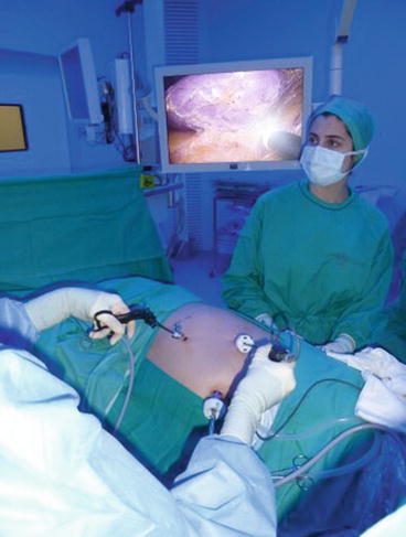
Fig. 7.2
Intraoperative placement of trocars in the case of reduced port splenectomy
7.4 Literature Review
7.4.1 Data Sources
A systematic search in PubMed (June 2013) was performed. The applied search strategy included the key words: (single incision OR single port) AND (laparoscopic OR laparoendoscopic OR robotic) AND splenectomy.
7.4.2 Case Reports of Single-Incision Laparoscopic Splenectomy
The main characteristics of the studies (demographics, medical history, clinical features of SILS performed, operative parameters, outcomes) are depicted in Table 7.1 [5–34]. A total of 81 patients were retrieved. The age ranged from 0.6 to 73 years. The majority of them were female (57 %). The median body mass index of the included patients was 23 kg/m2 (range: 18–36). Partial splenectomy was performed in two cases (2.5 %). The patients were positioned either in semi-lateral (62 %) or lateral position (38 %). The most frequent surgical approach was through the umbilicus (91 %), while the supraumbilical (6 %) or left upper quadrant (2.5 %) was also used. The median weight of the spleen was 446 g (125–590 g). Regarding the utilized port system, a multitrocar system (5–12-mm trocars) were applied in (54 %) patients, SILS® port in 27 %, TriPort® in six (8 %), glove ports in five (6 %), and GelPort® in four patients (5 %).
Table 7.1
Single-incision laparoscopic splenectomy. Literature review: single cases
Demographics | N (%) |
|---|---|
Age (year) | 23 (0.6–73) |
Male/female | 35/46 |
BMI (kg/m2) | 23 (18–36) |
Diagnosis | |
Splenomegaly | 33 (45 %) |
Idiopathic thrombocytopenic purpura | 23 (31 %) |
Immune thrombocytopenic purpura | 5 (7 %) |
Lymphomas | 5 (7 %) |
Splenic cysts | 4 (5 %) |
Traumatic rupture | 2 (3 %) |
Hydatic cyst | 1 (1 %) |
Multiple splenic abscesses | 1 (1 %) |
Spleen weight (g) | 446 (125–590) |
Operative parameters | |
Port system applied | |
2 or 3 single ports | 43 (54 %) |
SILS® port | 21 (27 %) |
TriPort® | 6 (8 %) |
Glove port | 5 (6 %) |
GelPort® | 4 (5 %) |
Size of incision (mm) | 22 (10–35) |
Outcomes | |
Operative time (min) | 125 (45–420) |
Blood loss (ml) 50 (10–450) | |
Conversion | 4 (5 %) |
Transfusion | 11 (14 %) |
Hospital stay (day) | 3 (1–9) |
Complications | 6 (7) |
Death | 0 |
Positive cosmesis | 42 (52 %) |
The median operative time was 125 min (45–420), while the median blood loss was 50 ml (10–450 ml). In 5 % a conversion was needed either to open or to multiport laparoscopic surgery, while transfusion was needed in 11 cases (14 %). The hospital stay ranged between 1 and 9 days. Complications related to splenectomy occurred in 7 % of patients. No death of patients was reported. Simultaneously, we identified five case series that included 58 SILS patients (Table 7.2) [4, 35–38]. Only in one study, the final cosmesis of the surgical wounds was evaluated with the difference being statistically significant in the single-incision group compared to the classical laparoscopic one. A similar systematic review has recently been published [39].
Table 7.2
Main characteristics and outcomes of patients after single-incision laparoscopic splenectomy, in published case series
Author/year | Study type | N | Age (yrs) | BMI (kg/m2) | Diagnosis | Device | Spleen weight (g) | Incision size | Duration (min) | Blood loss (ml) | Conversion (%) | Transfusion | Hosp. stay (day) | Complications | Cosmesis |
|---|---|---|---|---|---|---|---|---|---|---|---|---|---|---|---|
Boone 2013 [35] | Retrospective comparative | LS, 18/26 (69 %); SILS, 8/26 (31 %) | LS, 49 ± 17; SILS, 51 ± 22 p = 0.81
Related posts: Single-Incision Laparoscopic Appendectomy Single-Incision Laparoscopic Appendectomy
 Single Access Laparoscopic Cholecystectomy Single Access Laparoscopic Cholecystectomy
 Single-Access Robotic Surgery Single-Access Robotic Surgery
 Single-Access Laparoscopic Approach for Pancreatic Surgery Single-Access Laparoscopic Approach for Pancreatic Surgery
 Single-Access Laparoscopic Right Hemicolectomy Single-Access Laparoscopic Right Hemicolectomy
 Single-Access Laparoscopic Approach for Gastric Surgery (Hiatal Hernia Repair and Gastric Resections) Single-Access Laparoscopic Approach for Gastric Surgery (Hiatal Hernia Repair and Gastric Resections)
Stay updated, free articles. Join our Telegram channel
Full access? Get Clinical Tree
 Get Clinical Tree app for offline access

|