Fig. 12.1
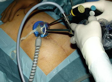
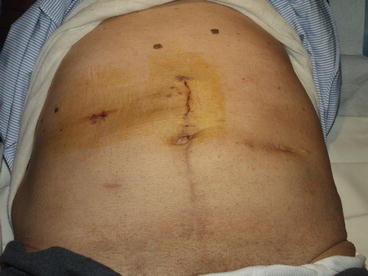
An umbilical or supraumbilical midline incision is generally performed for LESS liver resections, with the location of the incision varying depending on the body types and topography of the lesion
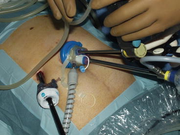
The majority of the authors describe the use of tools and the choice of a surgical technique very similar to the standard laparoscopic technique, with the difference that is performed through a single access [17–19]. Both 30° and linear 5–10 mm laparoscopes can be used to explore the peritoneal cavity. Then multiple instruments are simultaneously introduced in the abdominal cavity that generally consist of a tool for dissection along with one or more grasps or devices for aspiration (Fig. 12.2). The major technical difficulty in the execution of procedures through single-port technique is related to the parallel position that the devices mutually assume and the consequent loss of triangulation with the optical system, which limits considerably the possibilities of movement and the field of action. Therefore, some authors suggest the use of curved instruments specifically designed so as to obtain adequate exposure and triangulation, which can be an alternative to overcome this technical difficulty [20]. However, in the majority of cases, the use of straight laparoscopic instruments has been adopted, thus enabling the execution of these procedures without an increase in costs compared to traditional laparoscopy. Moreover, the adoption of standard laparoscopic instruments may shorten the surgeon’s learning curve in the acquisition of skills in the new single-port technique. As well as in open surgery, various methods and instruments for parenchymal transection during single-port liver resection are described, mostly corresponding to the methods adopted at individual centers for traditional laparoscopic liver resections. The methods for parenchymal transection generally preferred are ultrasound and radiofrequency-based devices [19–21]. As well as in open and laparoscopic surgery, the section of larger portal pedicles and hepatic branches can be managed through the use of articulating linear staplers. The single-port technique also allows the execution of intraoperative ultrasonography with a flexible laparoscopic ultrasound probe [14, 22]. The intraoperative ultrasound routinely performed before parenchymal transection allows the operator to identify precisely the location of the lesion and to plan the line of transection, which may be marked on the liver surface using diathermy. During parenchymal transection, the repeated use of intraoperative ultrasonography allows to ride along a transection plan oncologically adequate.


Fig. 12.2
Multiple instruments are simultaneously introduced in the abdominal cavity through the LESS device, generally consisting of a dissection tool along with one or more grasps or devices for aspiration
12.3 Discussion
The published series show the possibility to perform liver resections by single-port access without the need for conversion to traditional laparoscopy or open surgery. The conversion rate reported in literature ranges between 0 and 16.7 % [18, 20]. Adding further trocar to the device is not required to praxis but can be useful in case of need for better exposure of the operative field or for the management of complications. This occurrence may be necessary, in particular if using three-way single-port devices (Fig. 12.3) [19].


Fig. 12.3
Adding further trocar to the single-port device can be useful for better exposure of the operative field or for the management of intercurrent complications, in particular if using three-way single-port devices
Regarding intraoperative outcomes, the published series showed reduced blood loss and operating time and comparable to those obtained with traditional laparoscopic approach to hepatectomy (80.4–205 min, 2–500 mL) [14, 15]. As well as in traditional surgery, even for single-port hepatectomies, blood losses and operating times are closely related to the technique for transection, the occurrence of complications, the extent of resection, and the expertise of the operator, which implies certain variability with the completion of the learning curve.
The extraction of the surgical specimen can be carried out through the incision made for the insertion of the single-port device, after insertion in a suitable plastic bag retrieval, thus avoiding in most cases the need for additional abdominal wounds (Fig. 12.4) [20]. In case of benign neoplasms, the specimen can be broken up so as to avoid the need to expand the access for the extraction. In case of hepatectomy for malignant disease, for which the evaluation of the surgical margin and the integrity of the anatomical part are fundamental, it is possible to extend slightly the abdominal incision so as to remove the sample [11]. Therefore, it seems sensible to identify optimal candidates for the technique as patients affected by liver lesions smaller than 5 cm, so as not to dissolve the advantages related to the minimally invasive access. Some authors also recommend that the patient is not taller than 180 cm or suffering from obesity because in these cases the traditional laparoscopic instrumentation is not sufficiently long when inserted. Anyway, the single-port approach has already been reported to be safe and effective for bariatric surgery; therefore, obesity should not be considered an absolute contraindication in patients who are candidates to hepatectomy [23]. When considering the single-port approach, it should be rather argued that a thicker abdominal wall would lead to increased technical complexity because of instrument conflict at the fulcrum point. Another important criterion for the selection of patients seems to be the location of the lesions within the liver parenchyma. To be within reach of the laparoscopic instrumentation, hepatic lesions should be localized within laparoscopic segments (i.e., segments 2, 3, 4b, 5, 6), while lesions of the cranial and the first segment are not optimal candidates for a workable procedure. In this regard, at present, the best indication for the use of the single-port laparoscopy seems to be left lateral sectionectomy, because the transection line is in front of the device and significant triangulation of the instrument is not needed. In fact, the suspensory ligaments (round and falciform ligaments, left coronary and triangular ligaments) of the liver help the surgeon with the surgical field exposure, and in-line view is not a major issue to deal with [18]. The comparative analysis of a series of left lobectomy found that the single-port approach to liver resection is feasible, safe, and at least not inferior to the standard laparoscopic resection in terms of complications and clinical outcomes [24].


