Fig. 7.1
Table set-up for SRURS: 1 Guide wires, 2 Irrigation tubing, 3 Cystoscope and semirigid ureteroscope, 4 Light cord, 5 Video camera (clockwise from top left)
Room set up should mimic that of cystoscopy with ergonomic placement of video and fluoroscopic monitors. Normal saline is the preferred irrigation solution to limit adverse events as a result of potential systemic absorption during ureteroscopy. Pressure irrigation may improve visibility and most commonly is achieved with a pressure bag or hand-help pump syringe. A Level 1 System can also be used to control irrigation as well as to warm the irrigant to body temperature (Level NORMOFLO Irrigation Warming Set, Smiths Medical ASD, Rockland, MA, USA).
Cystoscopy is performed as an initial step to every ureteroscopy to rule out incidental bladder pathology and facilitate passage of the guide-wire into the ureter using fluoroscopy. We prefer to perform semirigid ureteroscopy along side a stiff guide wire such as an Amplatz Super Stiff™ wire (Boston Scientific, Natick, MA). A stiff wire has the potential benefit of straightening the course of the ureter, which is particularly useful in men with a large prostate and J-hook in the distal ureter or those with previous reimplants creating a distorted course of the distal ureter. This should however be exchanged after a floppy tipped guide-wire such as a Bentson™ wire (Cook Medical, Bloomington, IN) is placed into the ureter to limit the possibility of ureteral trauma/perforation.
Once access to the ureter is gained by passage of a guide-wire, this access should remain intact as a safety measure until the ureteroscopy procedure is completed. The guide-wire should be secured to the contralateral drape with a hemostat, which will prevent migration or inadvertent removal.
Passage of the semirigid ureteroscope should be performed with considerable caution as the tip of the scope can easily create false passages in the urethra or distal ureter. This is particularly true in the male urethra as the external sphincter and prostate can act as a fulcrum. It is imperative that the urethral and ureteral lumen be maintained in the center of the visual field to minimize the possibility of a false passage.
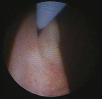
Upon entry into the bladder, the ureteral orifice is located and the scope is passed underneath the previously placed guide-wire. This maneuver facilitates passage of the scope into the distal ureter utilizing the guide-wire to open the ureteral orifice. This maneuver is demonstrated in Fig. 7.2. Occasionally, passing a second guidewire under direct vision through the working channel of the ureteroscope can facilitate traversing the ureteral orifice. Additionally, using pressurized irrigation may also help with passage of the scope into the ureteral orifice. However, when performing SRURS for the treatment of stones, the surgeon must be cognizant of the possibility of proximal migration of the stone as a result of the pressurized irrigation. If resistance is met during passage of the ureteroscope at any point during the procedure, the safety of proceeding should be weighed against the risk of potential complications.


Fig. 7.2
Attempting to insert semirigid ureteroscope into right ureteral orifice below guide wire
Although some caution against use of the semirigid ureteroscope for procedures proximal to the iliac vessels, advances in technology of the semirigid scopes can allow for safe passage into the proximal ureter in many patients. Obstacles to proximal ureteroscopy with the semirigid scope include large muscular habitus (presumably due to large psoas muscles), previous ureteral surgery, and obesity. If difficulties are encountered under these clinical settings, consideration of flexible ureteroscopy as an alternative must be exercised.
In the management of ureteral stones with semirigid ureteroscopy, there are a variety of treatment options. Among the instruments in the armamentarium of most urologists performing SRURS are various stone baskets, Holmium: YAG lasers of different diameters, as well as pneumatic and electrohydraulic (EHL) lithotripters. However, Holmium: YAG lasers have emerged as the standard of care in contemporary endoscopic lithotripsy. Reasons for laser lithotripsy are primarily related to improved efficacy and safety profiles.
Holmium lasers are not limited by stone composition, as they are effective in fragmenting all stone types. In addition, compared to EHL and pneumatic devices, the holmium laser produces smaller stone fragments during lithotripsy [12]. The holmium laser safety profile is significantly better than EHL as the laser energy is absorbed in the fluid medium of ureteroscopy and thus can safely be activated at a distance of 0.5–9 mm without the risk of ureteral injury or perforation [13].
Laser lithotripsy may be performed utilizing two different techniques depending on the stone size and characteristics. For the smaller stone, it is beneficial to “paint” or “dust” the stone with the laser fiber, essentially removing the outer borders of the stone as dust fragments. This can be done to completion, or if preferred, the stone may be reduced in size using this technique until amenable to stone basketing the fragments of the stone. In the setting of larger stones or impacted ureteral stones, the ureteral stone is typically fragmented into several pieces by using the laser to split the stones into halves. Once this is achieved, either the stones may be basketed or further fragmented using the “paint” technique.
There are a number of stone retrieval devices available for removal of stones or stone fragments during ureteroscopy. Many of these devices are Nitinol-based stone baskets, which are our preferred stone retrieval device. There are many variations of these devices, which are primarily tipless baskets or grasping devices, which combine the abilities of conventional stone baskets with those of three-prong graspers. These stone retrieval devices are discussed in more detail elsewhere in this book.
Whether it is necessary to leave a stent following semirigid ureteroscopy is a matter of some debate. At our institution, this decision is determined by individual surgeon preference, and characteristics of the patient and ease of the procedure weigh heavily on the ultimate decision to use a ureteral stent. There is level one evidence to support that after uncomplicated semirigid ureteroscopy, there is no benefit to ureteral stent placement. In fact, much of the literature demonstrates an increased incidence and severity of postoperative pain, increased analgesic use, and bothersome voiding symptoms in those patients who are stented following ureteroscopy [14, 15].
Tips and Tricks of Semirigid Ureteroscopy
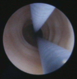
In the event of difficult access to the ureter, there are a number of potential tricks that may aid in the passage of the ureteroscope [16]. One such technique involves the use of an additional guide-wire through a working channel of the ureteroscope. This “railroad” technique may enable the ureteroscope to be more precisely guided into the distal ureter and facilitate passage of the scope as seen in Fig. 7.3. This may be of particular use when manipulating the ureteroscope proximal to the iliac vessels, and may overcome some patient-specific anatomical challenges such as the large psoas muscle mentioned previously.


Fig. 7.3
The railroad technique: one guide wire is seen adjacent to the SRURs. The second guide wire is inserted through the working channel of the ureteroscope, mimicking a “railroad track”
Another technique, particularly in females and children, is the use of serial fascial dilators up to 10Fr passed over a guide-wire under fluoroscopic guidance in order to dilate the ureteral orifice. The fascial dilators are inexpensive and have a good balance of tip flexibility as well as rigidity to safely dilate the ureteral orifice. In the male patient, it is our experience that the use of a balloon dilator is the preferred and safest method for dilating the ureteral orifice. This can be done either under fluoroscopy, direct vision, or a combination of the two. If either of these techniques does not allow passage of the scope, one may consider passage of a ureteral stent to allow for passive dilation and return for ureteroscopy at a later date.
Occasionally poor visualization is an issue when performing SRURS. In this event, any instrument within the working channel of the ureteroscope should be removed to identify this as an impediment to the inflow of irrigation. If this is not the source of the poor visibility, ensure that the scope is not bent using fluoroscopy, which may limit the irrigation potential. Irrigation with the use of a pressurized bag may be utilized to help improve visibility within the ureter, and this practice is our preferred method during ureteroscopy. Alternatively, an assistant may use the Single Action Pumping System™ (Boston Scientific, Natick, MA). This gives the surgeon control of the irrigation pressure and can be used when needed. When utilizing pressurized irrigation, the surgeon should be cognizant of the potential for ureteral stone migration as well as pyelovenous backflow, and therefore, the least amount of pressure necessary should be employed.
In the event of impacted stones, it may be difficult to gain access to the more proximal ureteral segment with the guide-wire. In this circumstance, our algorithm typically involves passage of a 5Fr ureteral catheter just distal to the stone. We then use an angled GlideWire to gently attempt traversing the impacted stone. If this is unsuccessful, one may inject some viscous lidocaine through the ureteral catheter in an attempt to lubricate the ureter in order to facilitate passage of the guide-wire.
Occasionally a stone may be grasped using a ureteral stone basket device without the ability to subsequently remove the stone or dislodge the stone from the basket. In this circumstance, the surgeon should disassemble the basket extracorporeally, which usually allows for removal of the basket after dislodging the stone from the basket. In the event that this is unsuccessful, the disassembled basket should be left within the ureter and the scope removed carefully. Subsequently, the semirigid ureteroscope may be passed alongside the basket and a laser fiber can be used to fragment the stone lodged within the basket. This should facilitate removal of the stone basketing device and hopefully the stone fragments.
Outcomes
Efficacy of semirigid ureteroscopy is measured by complete treatment of urolithiasis in the absence of adverse events. Non-uniform reporting plagues the urologic literature on outcomes with ureteroscopy, and thus conclusions drawn from the available series should be interpreted with some caution or at least awareness of the considerable heterogeneity of procedure and patient characteristics. In addition, the success of ureteroscopy varies considerably with respect to stone size and location, stone composition, type of ureteroscope used (semirigid versus flexible or a combination), lithotripter (laser, electrohydraulic, pneumatic), and surgeon experience. We discuss the available stone-free rates reported in contemporary literature.
In 791 semirigid procedures, El-Nahas et al. achieved a stone-free rate (SFR) of 87 % after a single operation for ureteral stones. Using a multivariate analysis, proximal ureteral location, stone impaction, stone width, and surgeon experience were independent predictors of unfavorable results (defined as residual stone or complication with the procedure) [17]. In a prospective, randomized trial of shock wave lithotripsy versus ureteroscopy for distal ureteral calculi, 29 of 32 patients in the semirigid ureteroscopy cohort were rendered stone free (90.6 %), with the remaining three patients being asymptomatic but without radiographic assessment of stone treatment efficacy [18]. Using a pneumatic lithotripter and semirigid ureteroscope, Aghamir et al. found an overall SFR of 89.5 % in 362 procedures with a mean stone size of 10.4 mm. However, the analysis included distal, mid, and proximal ureteral stones of all sizes with significant variation of success based on size and location, with the highest success rates being seen with distal stones less than 10 mm (97.4 %) [19]. Similarly, Sozen et al. reported on 500 patients undergoing SRURS for ureteral stones regardless of location. They quote an overall SFR of 94.6 %, with improved results with smaller stones (<10 mm 97.1 % versus >10 mm 83.7 % SFR) [20].
Although SFRs may be enhanced with the use of flexible ureteroscopes, Wu et al. demonstrated the feasibility of SRURS alone in the treatment of proximal ureteral stones, with a SFR of 83.2 %. When these stones were broken down into <10 mm and >10 mm, SFRs were 91.1 and 76.8 % respectively [21]. Using semirigid ureteroscopy for larger proximal ureteral stones (mean 18.5 mm), SFR was diminished considerably to 35 % demonstrating the impact of patient characteristics on successful outcomes [22].
When pooling the results of semirigid and flexible ureteroscopy, there is generally an improvement in reported overall SFR. With a mean stone size of 11.3 mm in 598 patients, use of SRURS below the iliac vessels and flexible ureteroscopy proximal to this, there was an overall SFR of 97 % in one series [23]. In a similar study focused on only proximal ureteral stones, 32 patients with a mean stone size of 8.3 mm were rendered stone-free in 96.8 % of cases using both flexible and semirigid ureteroscopy [24].
The Clinical Research Office of the Endourological Society (CROES) prospectively collected data on 9,681 patients undergoing ureteroscopic stone treatment in the Ureteroscopy Global Study. Stone free rates for distal, mid, and proximal ureteral stones were 94.2, 89.4, and 84.5 % respectively [25]. In a similar collective effort, the European Association of Urology and American Urological Association updated their guidelines on the management of ureteral calculi. Using pooled data of all ureteroscopy techniques via a meta-analysis, overall SFRs (independent of stone size) for distal, mid and proximal ureteral stones were 94, 86, and 81 % respectively [5]. Although success may vary based on stone size, location and experience among other parameters, semirigid ureteroscopy offers excellent stone free rates when used in the treatment of ureteral calculi. A summary of the above-mentioned studies is shown in Table 7.1.
Table 7.1
Stone-free rates in previously published series
Series
Stay updated, free articles. Join our Telegram channel
Full access? Get Clinical Tree
 Get Clinical Tree app for offline access

|
|---|


