Scrotal Swellings
Angela Cottrell
Benign swellings
Epididymal cyst
An epididymal cyst is a cystic swelling of the epididymis. Clinically, the patient may complain of a swelling in the scrotum that gets larger over time. Examination of an epididymal cyst will reveal a swelling that you can ‘get above’, which is separate from the testis. When transilluminating the cyst its appearance has a classical description of a ‘Chinese lantern’. If the patient has minimal symptoms, conservative management is appropriate. Aspiration is not appropriate as the cysts are frequently multiloculated. Surgical excision is appropriate if symptomatic (8.1).
Hydrocoele
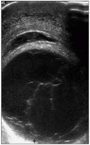
A hydrocoele is an abnormal collection of fluid between the visceral and parietal layers of the tunica vaginalis that surrounds the testis (8.2). It is the most common benign testicular mass, affecting 1% of males. The tunica vaginalis produces approximately 0.5 ml of fluid a day and the fluid accumulates due to an imbalance of production and absorption.
Causes of hydrocoele
Congenital: patent processus vaginalis.
Acquired: primary (idiopathic); secondary (i.e. trauma, infection, tumour).
The patient may give a history of a swelling in the scrotum. On examination of the scrotal swelling it is not usually possible to palpate the testis through the hydrocoele fluid collection. The surface of the swelling is smooth. It is possible to ‘get above’ the mass and it transilluminates.
Simple aspiration of a hydrocoele may achieve symptomatic relief of a tense hydrocoele; however, this is not definitive treatment as the fluid is likely to re-accumulate. Surgical intervention may include Lord’s plication technique or Jaboulay procedure involving excision of the hydrocoele sac. The hydrocoele fluid is then absorbed by scrotal lymphatics. In a child, the patent processus vaginalis may be ligated.
 8.1 Ultrasound scan of epididymal cyst. |
Varicocoele
A varicocoele is an abnormal dilation of the veins of the pampiniform plexus, which is present in 15% of men. The
patient may complain of a scrotal swelling and discomfort or dragging sensation in the scrotum. On examination it is possible to get above the mass. It is not possible to transilluminate the mass and characteristically it feels like a ‘bag of worms’. It increases in size when standing.
patient may complain of a scrotal swelling and discomfort or dragging sensation in the scrotum. On examination it is possible to get above the mass. It is not possible to transilluminate the mass and characteristically it feels like a ‘bag of worms’. It increases in size when standing.
 8.2 Ultrasound showing hydrocoele. |
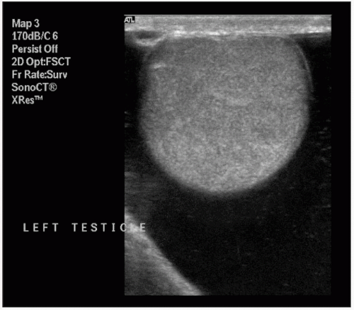
Varicocoeles are more common on the left side (90%) than the right due to the drainage of the left testicular vein into the left renal vein at an angle of 90°. Varicocoeles may arise secondary to a left-sided renal mass extending into the renal vein and causing back pressure on the testicular vein. It is therefore necessary to perform an ultrasound scan of the renal tract once a varicocoele has been identified to exclude renal pathology (8.3).
Acute epididymo-orchitis
Acute swelling of the epididymis or testis is most commonly infective in aetiology. Pathogens may be viral (e.g. mumps, orchitis) or bacterial. Pathogens usually arise from the bladder or urethra, however, and may rarely be secondary to systemic illness such as tuberculosis. In younger men, the most common pathogens are those from sexual transmission including Chlamydia trachomatis or Neisseria gonorrhoea. In older men, urinary tract pathogens are more frequent and bladder outlet obstruction may increase the risk of infection.
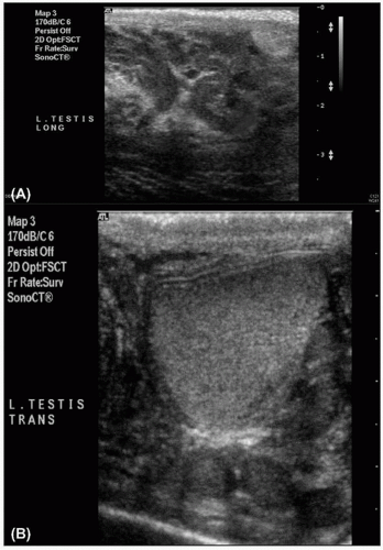 8.3 Ultrasound scan showing left varicocoele: (A) longitudinal and (B) transverse view. |
The man may present with a short onset (usually) unilateral testicular pain and may have a history of recent sexual activity. Examination may reveal a hot, tender testis or epididymis and the overlying skin may be erythematous (8.6).
Related posts:


Stay updated, free articles. Join our Telegram channel

Full access? Get Clinical Tree


