1. WG1, Handling of Radical Prostatectomy Specimens. R Montironi (Chair), H Samaratunga, L True
2. WG2, pT2 substaging and tumour volume in prostatectomy specimens. T Van der Kwast (Chair), M Amin, A Billis
3. WG3, Extraprostatic extension, lymphovascular invasion and locally advanced disease. PA Humphrey (Chair), C Magi-Galluzzi, AJ Evans
4. WG4, Seminal Vesicle and lymph node sampling. D Berney (Chair), T Wheeler, D Grignon
5. WG5, Surgical margins in radical prostatectomy specimens. J Epstein (Chair), L Cheng, P Hoon Tan
Many recommendations of this consensus conference have already been incorporated into international guidelines, including the recent College of American Pathologists protocol and checklist for reporting adenocarcinoma of the prostate and the structured reporting protocol for prostatic carcinoma from the Royal College of Pathologists of Australasia [10, 11].
In response to the question relating to how much of the prostate should be blocked, >60 % of conference participants supported complete embedding, whereas >60 % also supported partial embedding. This apparent contradiction arose as several respondents selected both options depending on the situation. In view of this, it was concluded that both methods were considered acceptable. Pathologists have to balance the extra expense and time involved in processing entire specimens against the risk of missing important prognostic parameters and decide whether partial or complete embedding should be performed. There was consensus that if partial embedding is performed, a specific protocol should be followed and the methodology should be documented in the pathology report [3].
From the survey, a majority of respondents reported using standard blocks and only 16 % reported the use of whole mounts, for at least some slices. A minority reported using both methods. On discussion at the consensus conference, it was considered that both standard blocks and whole mounts were acceptable for examination of radical prostatectomy specimens, although no ballot was taken on this point [3].
4.3 Total Versus Partial Embedding
For the pathologist, the safest method to avoid undersampling of cancer is evidently that the entire prostate is submitted. In some institutions, partial sampling is practiced. This requires that the pathologist adheres to a strict protocol, which may be somewhat cumbersome.
In 1994, a report on how prostate specimens were examined by American pathologists showed that only 12 % of pathologists embedded the entire prostate [12]. Since then the proportion of laboratories that use partial embedding has decreased. In a recent ENUP survey among 217 European pathologists from 15 countries, only 10.8 % used partial embedding routinely [13]. In some European countries, total embedding is even mandatory, according to national guidelines.
The recent study by Dr. Vainer et al. [14] analyses 238 RPS to determine whether significant prognostic information is lost when a partial sampling approach with standard cassettes is adopted, compared with total embedding. In their study, upon arriving at the pathology department, the prostate is partly divided by a cut in the mid-sagittal plane through the anterior surface, separating the two lobes for optimal fixation. The gland is then fixed for an additional 20 h in formic acid and 24 h in 4 % buffered formalin. The gross examination includes measurement in three dimensions, weighing the prostate after removal of the seminal vesicles, and separating the left from the right lobe after inking the anterior and the posterior halves with two different colours. Apical and basal slices of 5–10 mm, depending on the total size of the RPS, are cut horizontally, subsequently sliced parasagittally, and placed in cassettes with often more than one section per cassette. The remaining part of the prostate is cut horizontally in approximately 3-mm thick slices and placed in standard cassettes, ensuring laterality. Large slices are divided to fit standard cassettes. Finally, sections from the seminal vesicles (as a minimum the apex and a cross-section) are embedded. Post-fixation in 4 % formalin and embedding in paraffin are followed by 4-μm sectioning and staining with haematoxylin and eosin (number of cassettes/total slides: 18 to 76). For the purpose of the study, glass slides from every second horizontal slice are withheld (number of slides initially removed: 3 to 26, i.e. 29.9 %). The remaining slides are evaluated microscopically.
According to this group of researchers, such an approach decreases the laboratory workload by 30 %, and at the same time little information is lost with this procedure, overlooking features significant for the postoperative treatment in only 1.2 %. They conclude that partial embedding is acceptable for valid histopathological assessment.
The findings reported by Dr. Vainer et al. [14] are slightly better than those reported by others. Hall et al. [1] showed that by submitting only gross stage B cancer along with standard sections of the proximal and distal margins, the base of seminal vesicles and the most apical section (next to distal margin), 96 % of positive surgical margins and 91 % of instances of extraprostatic extension were detected, as compared with identification by complete microscopic examination. In the study by Cohen et al. [15] involving patients with clinical stage B carcinoma, each gland was serially sectioned with sections mounted whole on oversized glass slides. Using only alternate sections, there was a 15 % false-negative rate for extraprostatic extension. In a study by Sehdev et al . [4], cT1c tumours with one or more adverse pathological findings, such as Gleason score 7 or more, positive margins and extraprostatic extension, were compared using ten different sampling techniques. The optimal method consisted of embedding every posterior section and one mid-anterior section from the right and left sides of the gland. If either of the anterior sections had sizable tumour, all anterior slices were blocked in a second step. This method detected 98 % of tumours with Gleason score 7 or more, 100 % of positive margins and 96 % of cases with extraprostatic extension, through examination of a mean number of 27 slides. It was also shown that sampling of sections ipsilateral to a previously positive needle biopsy detected 92 % of Gleason score 7 or greater cancers, 93 % of positive margins and 85 % instances of extraprostatic extension, from a mean number of 17 slides.
4.4 The Ancona (Italy) Approach to the Evaluation of the Radical Prostatectomies
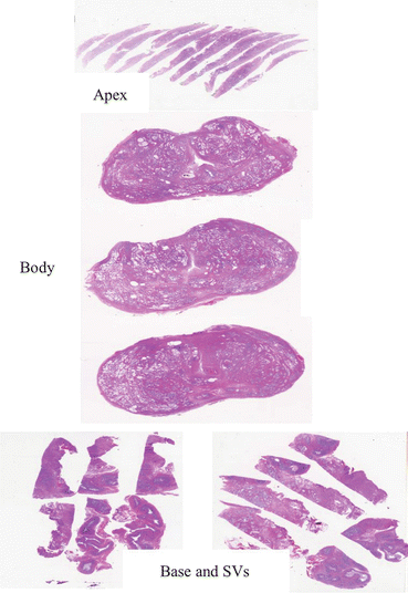
In the last few years, 3,000 RPS have been totally embedded and examined with the whole mount technique at the Section of Pathological Anatomy of the Polytechnic University of the Marche Region and United Hospitals, Ancona, Italy (Fig. 4.1) [2, 5].


Fig. 4.1
Prostate gland and seminal vesicles examined with the whole mount technique
The prostate is received fresh from the operating room. Its weight without the seminal vesicles and all three dimensions [apical to basal (vertical), left to right (transverse) and anterior to posterior (sagittal)] are recorded, the latter used for prostate volume calculation. To enhance fixation, 20 ml 4 % buffered formalin is introduced into the prostate at multiple sites using a 23-G needle. To ensure homogenous fixation, the needle is inserted deeply and the solution injected while the needle is retracted slowly. The specimen is then covered with India ink and fixed for 24 h in 4 % neutral buffered formalin. After fixation, the apex and base (3-mm thick slices) are removed from each specimen and examined by the cone method. The prostate body is step-sectioned at 3-mm intervals perpendicular to the long axis (apical–basal) of the gland. For orientation, a cut with a surgical blade is made in the right part of each prostate slice. The seminal vesicles are cut into two halves (sandwich method) and processed in toto. The cut specimens are post-fixed for an additional 24 h in 4 % neutral buffered formalin and then dehydrated in graded alcohols, cleared in xylene, embedded in paraffin (the material is processed together with regular cassettes), and examined histologically as 5-μm thick whole mount haematoxylin and eosin (H&E) stained sections [2, 5].
The body of each prostate is represented with 3–6 whole mount slides, whereas the apex, base and seminal vesicles with 6–8 regular slides, totalling between 9 and 14 slides (In Dr. Vainer et al.’s study [14], up to 76 regular slides are needed to examine the whole prostate). The time needed to section each specimen with an ordinary delicatessen meat slicer is 15–20 min. The time taken by a technician to cut all the blocks of an individual case is 30–40 min. The time needed by the pathologist to report a case ranges from 40 to 60 min. Since the slides do not fit into the current staining machines, the slides are manually stained. The paraffin blocks and glass slides are stored in dedicated containers because of their large size. The comparison between Dr. Vainer et al.’s and our approach is presented in Table 4.2 [14].
Table 4.2
Comparison between Ancona experience and Dr. Vainer et al.’s study
Features | Ancona experience | Dr. Vainer et al.’s study |
|---|---|---|
Prostate weight and size (and volume) | Yes (yes) | Yes (not mentioned) |
Fixation enhancement | Formalin injection | Separating the two lobes |
Inking of the surface | One colour; orientation with a cut on the right | Two colours, anterior and posterior halves |
Pre-sectioning fixation (time) | 4% Buffered formalin (24 h) | Acid formic (20 h) and 4 % buffered formalin (24 h) |
Sectioning interval | 3 mm (Apex and base: 3 mm) | Approximately 3 mm (Apex and base: 5–10 mm) |
Sub-division of the slices of the prostate body | No (Whole mounts) | Yes, to fit standard cassettes |
Seminal vesicles | Sandwich method (all included) | As a minimum the apex and a cross-section |
Post-sectioning fixation (time) | 4 % Buffered formalin (24 h) | 4 % buffered formalin (not mentioned) |
No. of cassettes/total slides (% examined) | 9–14 (100 %) | 18–76 (70 %) |
Processing | As for regular size cassettes | Not mentioned |
Slide size (section thickness) | 7.5 cm by 5.0 cm (5 μm) | 7.5 cm by 2.5 cm (4 μm) |
Slide staining procedure | Manual | Not mentioned |
Slides with substandard sections, however with cancer still evaluable, were observed in 7 cases (0.23 % of RPS). Only in one case (0.03 %), the quality was so poor that the features could not be evaluated. An individual block had to be serially sectioned to visualize the entire inked surface in 15 cases (0.5 %). Immunohistochemistry (mainly the basal cell marker p63, racemase and Chromogranin A) was done, always successfully, in 30 cases (1 %), cutting from the whole mount section the part to be evaluated in 28, and using the whole mount section in the remaining two. A procedure was developed to search for residual cancer prostate cancer on pT0 radical prostatectomy after positive biopsy [16, 17]. When applied to 10 cases, a minute focus of cancer was successfully found in 8 [16].
Stay updated, free articles. Join our Telegram channel

Full access? Get Clinical Tree


