Distal ureteral reconstruction is increasingly being performed by minimally-invasive surgical techniques. The robotic surgical platform provides an additional modality for repairing distal ureteral defects with the associated benefits of a minimally-invasive approach. This article reviews and describes the technical aspects of robotic distal ureteral reconstruction. In addition to discussion of the operative technique, factors such as patient selection, preoperative and postoperative evaluation, and published outcomes are addressed.
Key points
- •
Advancements in robot-assisted laparoscopic surgery have led to the development of minimally invasive techniques for distal ureteral reconstruction.
- •
Enhanced visualization and range of motion with the robotic platform improves the feasibility of laparoscopic, minimally invasive distal ureteral reconstruction.
- •
Adjunctive procedures including vesicopsoas hitch and Boari flap are also technically feasible through a minimally invasive approach using the robotic platform.
- •
Initial outcomes of robotic distal ureteral reconstruction seem safe and effective, and compare well with traditional open approaches.
Introduction
The treatment of distal ureteral defects, including benign stricture disease, fistulae, and malignancy, often requires ureteral reconstruction to restore ureteral patency and normal renal drainage. Historically, distal ureteral reconstruction has been performed via open surgical intervention after failed initial endourologic management, with success rates quoted well over 90%. Minimally invasive techniques for distal ureteral reconstruction were initially described nearly 2 decades ago, with reports of the first laparoscopic ureteroureterostomy by Nezhat and colleagues in 1992 and laparoscopic ureteral reimplant by Ehrlich and Gershman in 1993. Since that time, technological advancements in operative laparoscopy, including use of the da Vinci robotic operating platform, has allowed urologists to expand the scope of minimally invasive treatment options for distal ureteral defects.
Laparoscopic surgical approaches offer inherent benefits compared with open options when considering factors such as blood loss, hospital stay, and postoperative cosmesis. Despite this, pelvic laparoscopy often presents significant technical difficulty, and previously was reserved for those with advanced training. Use of the da Vinci operating platform across the wide scope of urology has increased surgeon comfort with minimally invasive surgery and has provided other technical benefits including three-dimensional and magnified visualization, enhanced range of motion and dexterity, and improved ease of intracorporeal suturing. Robotic surgery seems to have its greatest benefit in procedures requiring fine operative movements, where range of motion is limited, and where visibility is impeded.
Several institutions have recently described their early experience with robotic distal ureteral reconstruction, including application of supportive techniques such as vesicopsoas hitch and Boari flap creation. In this article, these publications are reviewed and the technical aspects of distal ureteral reconstruction are described. In addition to discussion of the operative technique, it is imperative to review factors such as patient selection and preoperative and postoperative evaluation.
Patient selection
Patient selection is an essential factor for successful completion of a robotic distal ureteral reconstruction. As in planning for any procedure, patient comorbidity factors should be addressed and optimized before the operation. Factors such as age, body habitus and mobility, previous surgical history, and body mass index need to be considered.
Although technically feasible, robotic ureteral reconstruction in patients with morbid obesity may present additional operative challenges not encountered in the nonobese patient. Increased abdominal girth may inhibit the full range of motion of robotic arms and instruments and limit the full extension of the instruments into the pelvis. In these situations, longer trocars and instruments may assist with successful completion of the operation. Also, great care should be taken to prevent operative positioning injuries, with special attention given to padding all pressure points and areas of bony prominence.
Patients with significant past surgical histories including multiple intra-abdominal operations or use of abdominal mesh may prove to be less optimal candidates for robotic distal ureteral reconstruction. A history of previous extensive intra-abdominal surgeries is another relative contraindication to robotic ureteral reconstruction. Significant scarring and adhesion formation may impede the development of proper anatomic surgical planes. In addition, substantial adhesion formation may inhibit the ability to establish the operative domain and prolong operative times due to excessive adhesiolysis.
Preoperative evaluation
The overall preoperative evaluation may vary greatly from patient to patient given the etiology of distal ureteral pathologic conditions; however, proper imaging is a mainstay in patient evaluation and operative planning. The location of the pathologic lesion, the length of the affected ureteral segment, and the extent of the defect must be defined during preoperative planning as these factors may influence the type of repair. Patients often present with basic imaging that demonstrates obstruction, including axial imaging studies such as ultrasonography or computed tomography (CT) scans. Triple-phase CT scans provide particular benefit as delayed images demonstrate ureteral filling and associated defects or obstruction. Nuclear medicine radioisotope studies including Mag-3 Lasix renograms may be of benefit in assessing the degree of preoperative obstruction, and can also be used in postoperative follow-up to demonstrate the patency of the repair. A functional study, such as a Mag-3 renogram, also confirms adequate renal function before planning repair. Depending on the presentation, some patients may undergo initial management with ureteral stent placement or nephrostomy tube placement. Retrograde pyelography and antegrade nephrostograms, or a combination of the two (an aptly named up and down-o-gram), may also provide a dynamic view of the affected ureteral segment, and provide further information for operative planning.
Patients with a history of urothelial malignancy, or when an index of suspicion for malignancy exists, should undergo endoscopic evaluation via ureteroscopy with biopsy, brush biopsy, or ureteral washing. The imaging studies mentioned earlier, particularly a triple-phase contrast CT scan, generally provide sufficient information to determine whether preoperative ureteroscopy is necessary before repair.
In assessing the diseased ureteral segment, determination of the length of the potential defect to be bridged is paramount to planning the necessary repair and assisting in preoperative counseling. Defects of 2 to 3 cm may be managed with ureteroureterostomy, whereas defects of 4 to 5 cm may be better managed via ureteroneocystotomy. For defects measuring 6 to 10 cm, a vesicopsoas hitch may be more appropriate. A Boari flap or bladder advancement flap may be used for longer defects measuring 12 to 15 cm in length ( Table 1 ).
| Technique | Length of Ureteral Defect (cm) |
|---|---|
| Ureteroureterostomy | 2–3 |
| Ureteroneocystostomy | 4–5 |
| Vesicopsoas hitch | 6–10 |
| Boari flap | 12–15 |
Operative evaluation and procedural positioning
Before proceeding to the operating room for a robotic distal ureteral repair, medical evaluation and clearance from a multidisciplinary team are recommended, including the urologic primary surgical team, the anesthetic team, and any medical consultants if specific concerns need to be addressed. Our patients are administered a gentle preoperative bowel preparation consisting of magnesium citrate and a clear liquid diet the day before the procedure. Patients should also receive appropriate perioperative antibiotic prophylaxis in accordance with current guidelines, usually consisting of a first-generation cephalosporin or fluoroquinolone depending on allergy status. Sequential compression devices are placed before induction of the general anesthetic.
After induction, the patient is placed in a dorsal lithotomy position with the arms tucked at their sides. Great care should be taken to keep the arms in a slightly supinated, neutral position with appropriate padding to avoid potential median or ulnar nerve injury. An orogastric tube is placed per anesthesia for gastric decompression. Patients should be secured to the operating table via padding and tucking of arms, or using a surgical bean bag immobilizer. After appropriate positioning, a tilt test may be performed to ensure that the patient will tolerate a steep Trendelenberg position without anesthetic issues or change in positioning. Proper steps should also be taken to ensure access to nephrostomy tubes if present, as injection via the nephrostomy tube or clamping may induce hydroureter, subsequently aiding in ureteral identification, dissection, and transection. In addition, removal of the nephrostomy tube in the operating room may help to prevent malpositioning of any ureteral stents placed during the repair.
Some investigators advocate performing cystoscopy with retrograde placement of a ureteral catheter with or without a wire before the abdominal portion of the operation. Ureteral stenting is a necessary part of the procedure and aids in healing of the repair; our institution has favored placement of the ureteral stent via a wire passed through an assistant port with direct visualization of the ureteral stent placement during the repair. Depending on the nature of the stricture, retrograde stent placement may not be possible. After cystoscopy and ureteral catheter placement or before establishing pneumoperitoneum, a Foley catheter should be placed for bladder decompression. In cases where an associated vaginal defect exists such as ureteral-vaginal fistula, a sponge stick or EEA sizer inserted per vagina may assist with visualization and dissection. In cases where the distal ureteral segment is to be excised because of malignancy, endoscopic scoring of the mucosa surrounding the ureteral orifice has been advocated to facilitate removal of a bladder cuff at the time of resection.
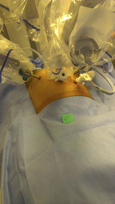
In addition to dorsal lithotomy positioning, patients are placed in a moderate to steep Trendelenberg position to allow the abdominal contents to fall out of the operative field as much as possible. Previous publications have indicated that tilting the table upward on the affected side, or airplaning the table, may also assist with preserving visualization within the operative field. The robotic surgical system is then brought toward the operative field between the patient’s legs when supine or over the ipsilateral iliac crest when in a tilted position ( Fig. 1 ).
Related posts:
 Urologic Trauma and Reconstruction
 Foreword
 Tips for Successful Open Surgical Reconstruction of Posterior Urethral Disruption Injuries
Primary Realignment of Pelvic Fracture Urethral Injuries
Skin Grafting of the Penis
Tips for Successful Open Surgical Reconstruction of Posterior Urethral Disruption Injuries
Primary Realignment of Pelvic Fracture Urethral Injuries
Skin Grafting of the Penis
 Reconstruction of Traumatic and Reoperative Anterior Urethral Strictures via Excisional Techniques
Stay updated, free articles. Join our Telegram channel

Full access? Get Clinical Tree


