Fig. 7.1
Port configuration. Three 5 mm ports through single-port device
The site for the single-port device and subsequent extraction is often times chosen to be right through the umbilical stalk. This hides the incision with the aim of better cosmesis. It is unclear in the literature if this can result in an increased incisional hernia rate. The incision size depends on the bulk of the colon and tumor, but typically starts around 3–4 cm. Alternatively, the port can be placed in the supraumbilical location, which is typical for a conventional laparoscopic approach.
Operative Steps (Table 7.1)
Table 7.1
Operative steps
Operative steps | Degree of technical difficulty (scale 1–10) |
|---|---|
1. Insertion of the single port and exploratory laparoscopy | 1 |
2. Identification and ligation of the ileocolic vessels | 5 (medial to lateral) |
4 (rollover technique) | |
3. Dissection of the retroperitoneal plane and identification of the duodenum | 5 (medial to lateral) |
4 (rollover technique) | |
4. Mobilization of the right colon and terminal ileum | 3 |
5. Mobilization of the proximal transverse colon and hepatic flexure | 5 (medial to lateral) |
4 (rollover technique) | |
6. Identification and ligation of the middle colic vessels | 6 (rollover technique) |
7. Extracorporeal anastomosis, closure, and reinspection | 4 |
The surgeon stands on the right side of the patient using two working instruments, which typically cross at the level of the fascia (Box 7.1). One hand is retracting the colon while the other is performing the dissection. Countertraction is achieved by gravity and anatomic fixation of the tissue such as the attachment of the colon to the peritoneum along the line of Toldt or the omentum to the stomach. The assistant stands away and to the right of the surgeon and is adjusting the camera to balance centering on the target and avoiding collision with the instruments.
Box 7.1 Tip
Keeping the target at the side of the camera’s field of vision avoids instrument collision.
Insertion of the Single Port and Exploratory Laparoscopy
The incision and initial access is very similar to a Hasson technique. The length of the skin incision can be usually less than the fascial incision size of about 3–4 cm. Once access into the peritoneum is achieved, the surrounding fascia is checked with a finger sweep to rule out any adhesions and the single-port device with preplaced trocars is inserted. The abdominal cavity is then explored in a standard fashion to rule out metastatic disease. Adhesiolysis can be performed (see Video 7.1).
Identification and Ligation of the Ileocolic Vessels
The surgeon’s right hand is using a bowel grasper and retracting the cecum or the mesentery close to the cecum towards the right abdominal wall. This allows visualizing the ileocolic pedicle. The left hand is using a dissecting instrument such as a bipolar vessel sealer or monopolar hook or scissors to isolate the ileocolic pedicle and start the medial to lateral dissection separating the colon mesentery from the retroperitoneum. As the instruments cross, maximum fulcrum effect is achieved. Once the duodenum is identified, the pedicle can than be divided (see Video 7.2). In the alternative approach (“rollover technique”), the ileum and small bowel mesentery is divided first, and the ileocolic pedicle can be visualized by continuously alternating between lateral colon mobilization and mesenteric division until the duodenum and the base of the pedicle are identified.
Dissection of the Retroperitoneal Plane and Identification of the Duodenum
After division of the ileocolic pedicle, the right hand continues to lift the cecum and the left hand dissects between the ascending colon mesentery and the retroperitoneum. The extent can be limited by the reduced ability to tent up the colon. During the alternative approach the dissection of this plane is from lateral and inferior.
Mobilization of the Right Colon and Terminal Ileum
The left hand now grasps the cecum with a bowel grasper and retracts medially and cephalad. The right hand is using an energy device or a monopolar hook or scissors to mobilize the colon along the line of Told and separating the remaining mesentery off the retroperitoneum. The instruments again cross to avoid instrument collision (see Video 7.3).
During the alternative rollover technique, this is the first step. The ileum is identified 10 cm proximal to the ileocecal valve, a mesenteric window is created, and the ileum divided using a laparoscopic stapler (see Video 7.4). The distal ileum is now tented up with the left hand in a medial and cephalad direction and the small bowel mesentery divided towards the base of the ileocolic pedicle. Once the division is close to the retroperitoneum, the cecum and colon mesentery is mobilized laterally for a section and the mesentery subsequently divided (see Video 7.5). The cecum and ascending colon “rolls over” medially and cephalad and the cycle is repeated until the duodenum and ileocolic pedicle is isolated.
Mobilization of the Proximal Transverse Colon and Hepatic Flexure
The right hand now retracts the transverse colon inferiorly to achieve adequate tension of the omentum. The left hand divides the omentum to enter the lesser sac and the hepatocolic attachments (see Video 7.6).
During the rollover technique the lateral to medial dissection is continued around the hepatic flexure with ongoing retraction of the ascending and transverse colon medially and then inferiorly to divide the omentum from the right lateral side.
Identification and Ligation of the Middle Colic Vessels
The division of the middle colic vessels is difficult from the inframesocolic approach because only one retracting instrument is available, and the transverse colon can often times not be lifted enough to have adequate exposure. The right branch can be divided though safely from superior and right lateral using the rollover technique.
Extracorporeal Anastomosis, Closure, and Reinspection
Once the dissection is completed, the specimen is extracted through the single-port incision site. The limiting factor is often times the bulk of mesentery and/or the size of the tumor (Box 7.2). With the rollover technique the small bowel mesentery and terminal ileum are already divided intracorporeally and the specimen can be therefore extracted easier through a smaller fascial incision. The divided distal ileum is grasped and the specimen extracted as a tube and not a loop (see Fig. 7.2). The extracorporeal anastomosis is fashioned in a standard way and the abdomen reinspected prior to closure (see Video 7.7).
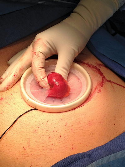
Fig. 7.2
Ileal extraction
Box 7.2 Caveat
When extracting the divided proximal ileum, ensure that the mesentery is not twisted for an extracorporeal anastomosis.
Approaches
Medial to Lateral Approach
A medial to lateral approach is the most common utilized approach in a conventional laparoscopic right hemicolectomy is a logic choice. This follows steps 1–7. It should be emphasized that extra care should be taken for safe vessel ligation of the ileocolic pedicle as intraoperative bleeding is difficult to control with availability of only two working instruments and limited mobility.
Rollover Technique or Modified Lateral to Medial and Inferior Approach
A very safe and easy alternative to the standardized medial to lateral approach is a modification and combination of the lateral to medial and inferior approach (Box 7.3). It specifically addresses the often times difficult dissection of the ileocolic pedicle, which is the first step of the medial to lateral approach.
Box 7.3 Tip
The rollover technique can be very useful for conventional laparoscopic right hemicolectomies in the morbidly obese with a very short and thick colon mesentery and ileocolic pedicle.
This alternative is also called the “rollover technique” because the cecum, ascending colon, and hepatic flexure are progressively rolled over in a clockwise fashion during different parts of dissection. This allows having adequate tissue exposure with traction through the instrument and countertraction through the attachment of the colon to the peritoneum along the line of Told and the hepatocolic ligament, mesenteric attachments to the root, and omental attachments to the stomach. The approach follows steps 1, alternating 3 and 4, 2, 5, 6, and 7. It should be emphasized that the procedure starts with creating a mesenteric window and dividing the terminal ileum first, followed by continuous division of the small bowel, and then colon mesentery alternating with dissection of the mesentery of the retroperitoneum and later duodenum.
Related posts:
 Right Hemicolectomy and Ileocecectomy: Robotic Intracorporeal Anastomosis
 Sigmoid Colectomy and Left Hemicolectomy: Hand-Assisted Laparoscopic Approach
 Right Hemicolectomy and Ileocecectomy: Single-Port Robotic Approach
 Operating Room Setup and General Techniques in Minimal Invasive Colorectal Surgery
 Proctectomy: Total Robotic Approach
 Sigmoid Colectomy and Left Hemicolectomy: Single-Port Laparoscopic Approach
Stay updated, free articles. Join our Telegram channel

Full access? Get Clinical Tree


