Renal Infections
Sameh K. Morcos MD, FRCS, FRCR
Case 6-1
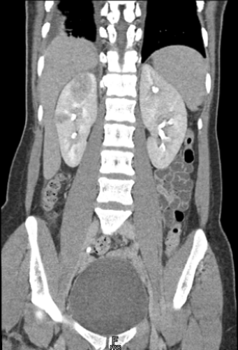
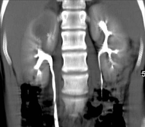
History: 26-year-old female with right flank pain, fever, nausea, and positive urine culture.
Findings: Coronal reformatted image from a CT urogram demonstrates a focal round low-attenuation area in the upper pole as well as a smaller area in the mid right kidney.
Diagnosis: Multifocal acute bacterial pyelonephritis
Discussion: Acute pyelonephritis is used to refer to an acute inflammatory infectious process affecting the renal parenchyma. Most affected patients are young or middle-aged women. Infection usually begins in the bladder and ascends to involve the ureters, intrarenal collecting systems, and then the kidneys. The most common offending organism is Escherichia coli (E. coli). On images obtained at CT urography, acute pyelonephritis may produce one or more focal, round areas of low attenuation, as in this case, or diffuse renal enlargement. Portions of the kidney may also show alternating linear areas of increased and decreased attenuation, creating a striated nephrogram. Treatment with antibiotics is usually effective; however, in some patients, antibiotic therapy may be needed for 6 to 8 weeks. (Case courtesy of Satomi Kawamoto, MD, Johns Hopkins Medical Institutions, Baltimore, MD.)
Case 6-2
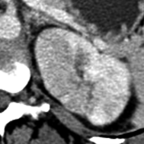
History: 33-year-old female with a history of tuberculosis presents with fever, left flank pain, and elevated white blood cell count.
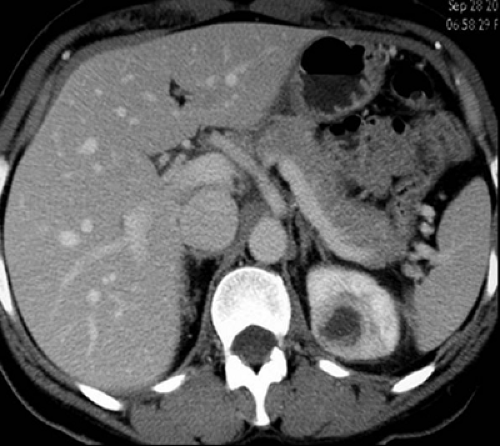
Findings: Axial image of the left kidney during the nephrographic phase obtained as part of a CT urogram shows an area of ill-defined, low density in the renal parenchyma.
Diagnosis: Acute pyelonephritis
Discussion: In patients with acute pyelonephritis, CT urography may demonstrate focal areas of low attenuation on nephrographic phase images. These areas of abnormally decreased enhancement may be patchy, round, or wedge shaped.
In comparison, delayed excretory phase images may reveal increased accumulation of contrast material in or adjacent to the abnormal previously hypodense parenchyma. CT urography in this case showed no signs of tuberculosis; the detected changes were compatible with the subsequently clinically established diagnosis of acute bacterial pyelonephritis. Tuberculosis typically produces more severe changes in the kidneys, including well-defined areas of low attenuation in the kidneys, renal parenchymal calcifications, and renal collecting system distortion and dilatation (see Cases 6-12 and 6-13). (Case courtesy of Stuart G. Silverman, MD, Brigham and Women’s Hospital, Boston, MA.)
Case 6-3
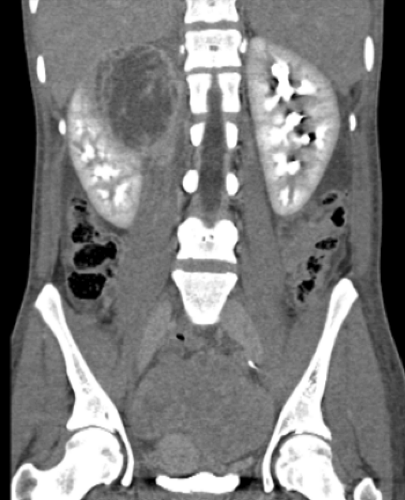
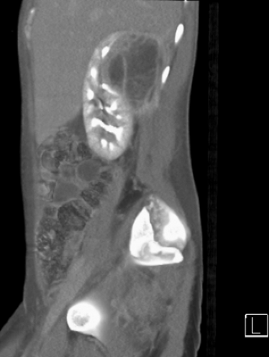
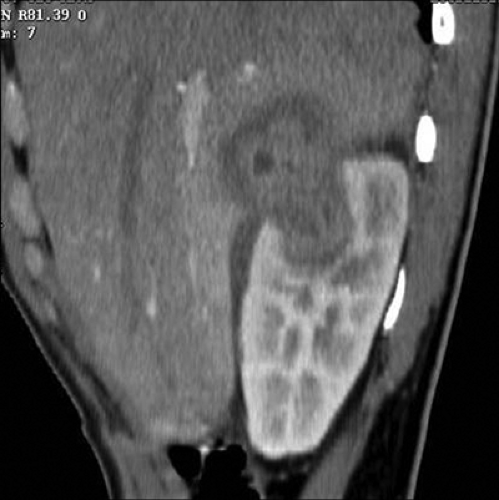
History: 30-year-old female with history of intravenous drug abuse presents with chills, right flank pain, and increased urinary frequency.
Findings: Coronal (A) and sagittal (B) enhanced images from a CT urogram show a 7-cm mass in the upper pole of the right kidney. The mass is predominantly hypodense with multiple enhancing septations, and it contains an irregularly thickened and enhancing wall.
Diagnosis: Renal abscess
Discussion: Although most acute renal infections resolve with antibiotic therapy, in some patients, particularly those who are immunosuppressed, renal abscesses may develop. Renal abscesses form in most patients as a result of inadequate treatment of an ascending infection, typically with E. coli. Intravenous drug abusers may develop renal abscesses as a result of hematogenous seeding. In these patients, Staphylococcus, Streptococcus, and Enterobacter species are more common than E. coli. On images obtained at CT urography, renal abscesses may have an appearance similar to that of a benign complicated renal cyst. In the appropriate setting, an abscess may be diagnosed when a cystic mass demonstrates a thickened, inflamed wall surrounding a hypodense fluid collection. Perinephric stranding, found commonly in the setting of inflammatory processes, is usually present and an important clue to the diagnosis. In this case, the abscess was due to Staphylococcus aureus and treated successfully with percutaneous CT-guided needle aspiration and catheter drainage. (Case courtesy of Satomi Kawamoto, MD, Johns Hopkins Medical Institutions, Baltimore, MD.)
Case 6-4
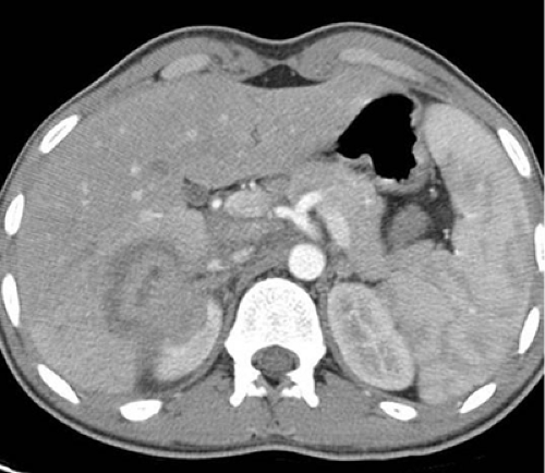
History: 18-year-old male with right loin pain and recurrent fevers for 4 weeks despite antibiotics.
Findings: Axial (A) and sagittal (B) corticomedullary phase images from a CT urogram show a 4.5-cm mass involving the right kidney and a peripheral portion of adjacent liver. The mass has a thick enhancing wall and a nonenhancing, hypodense central portion. Early excretory phase images (C, D) demonstrate hyperenhancement of the adjacent renal parenchyma. Delayed (30-minute), thick coronal slab maximum intensity projection image (E) shows that the mass fills with contrast medium.
Diagnosis: Renal abscess
Discussion: If left untreated, renal abscesses may enlarge and spread locally. Renal abscesses may penetrate through the renal capsule into the perinephric space and result in perinephric abscess formation. Although uncommon, renal abscesses may also involve adjacent organs, as in this case, in which a portion of liver was involved. Finally, as this case demonstrates, a renal abscess may excavate into the collecting system. As a result, the abscess may be opacified with excreted contrast material on delayed excretory phase images. Urine culture is typically positive in patients with ascending urinary tract infections; however, it may be negative in patients who develop renal abscesses due to hematogenous seeding. The offending organism was not cultured in this case. (Case courtesy of Tarek El Diasty, MD, Mansoura University, Mansoura, Egypt.)
Case 6-5
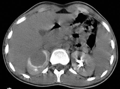
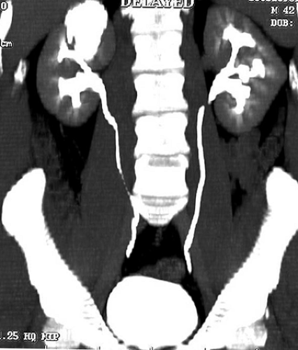
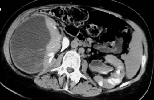
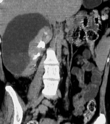
History: 32-year-old paraplegic male who developed a urinary tract infection and right flank pain despite prophylactic antibiotics.
Findings: Axial (A) and coronal (B) excretory phase images from a CT urogram demonstrate a large, low-attenuation collection along the lateral aspect of the right kidney; the renal parenchyma is severely compressed. There is bilateral moderate caliectasis and multifocal renal parenchymal loss on the left, likely due to chronic pyelonephritis or reflux nephropathy.
Diagnosis: Subcapsular renal abscess
Discussion: Infected subcapsular fluid collections are an uncommon sequela of renal infection. They are best treated with percutaneous catheter drainage. CT urography can be used to demonstrate the subcapsular location of the fluid, determine its extent, and help plan the percutaneous drainage procedure. In this case, percutaneous catheter drainage yielded purulent material, although no organisms were identified (possibly because the patient was already being treated with antibiotics). The patient recovered following percutaneous drainage and continued treatment with broad-spectrum antibiotics.
Case 6-6
History: 42-year-old female with pyuria, fever, and flank pain.
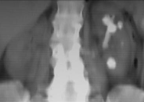
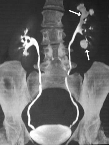
Findings: Unenhanced coronal average intensity projection image (A) from a CT urogram shows a large branch calculus in the upper pole of the left kidney, a smaller round calculus in the lower pole, and several additional small calculi in the mid kidney. Excretory phase volume-rendered image (B) demonstrates excretion into both renal collecting systems. Calices in the left upper and lower poles are blunted. The calculi in the upper (superior arrow) and lower (inferior arrow) poles can still be seen because they are lower in attenuation than the excreted contrast media. The calcification in the lower pole is located in a caliceal diverticulum. An axial image (C) shows that there is a 3-cm heterogeneous mass in the upper pole of the left kidney, adjacent to the large branch calculus. This mass contains a hypodense center and an evenly thickened wall.
Diagnosis: Left nephrolithiasis and left renal abscess
Discussion: Although some types of urinary tract calculi (e.g., calcium phosphate and magnesium ammonium phosphate stones) may form in chronically infected alkaline urine, urinary tract infection and abscess formation can also be a complication of untreated stone disease, as in this case. When urinary tract infections and stones are concurrent, the infection will often not be treated adequately until the stone, which can serve as an ongoing nidus for infection, has been removed. Many renal abscesses require drainage; however, when the abscesses are small, long-term antibiotic therapy may suffice. (Case courtesy of Richard Cohan, MD, and Elaine Caoili, MD, University of Michigan, Ann Arbor, MI.)
Stay updated, free articles. Join our Telegram channel

Full access? Get Clinical Tree


