Fig. 3.1
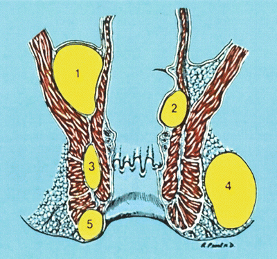
(Left) Anal glands opening in the anal crypts and branching in intersphincteric space. (Right) Extension of abscess to adjacent spaces. 1. Submucosal, 2. High Intermuscular, 3. Supralevator, 4. Ischiorectal, 5. Perianal. Provided courtesy of Russell K. Pearl, M.D., FACS, Department of Surgery, UIC
Aside from cryptoglandular origin, the other causes of anorectal suppuration may result from the downward spread of pelvic infection from appendicitis, diverticulitis, and gynecologic sepsis resulting in supralevator abscess. This in turn may track caudad through the levators into the ischiorectal space. Crohn’s disease of mid and low rectum as well as the anorectum, by its transmural pathologic nature may extend to perirectal or perianal space as an abscess fistula. Similarly, ingested chicken and fish bones as well as swallowed toothpicks may puncture the wall of the rectum resulting in significant sepsis. External penetrating injuries (stab, gunshot, shot gun wounds) may result in an overt injury though at times more minor or subtle injuries gradually proceed to full-blown perirectal sepsis. Perforation of low rectal cancers may also present with large ischiorectal abscesses. It is important to biopsy the abscess well in order not to miss the underlying cancer. Specific infections such as tuberculosis have been covered in Chap. 19. Fungal infections, especially actinomycosis and oxyuris vermicularis infestation, have also been known to produce abscess fistulas in adults and children.
Clinical Manifestation
The clinical picture of anorectal abscess depends on the level (height) of the abscess. If an imaginary line is drawn across the anorectal ring, supralevator and high intermuscular abscess are located above this level. These will often manifest with systemic symptoms of fever, toxicity, etc., but are not always associated with pain and other local symptoms. Their diagnosis often relies on a thorough rectal examination which can be done under anesthesia in the operating room if needed. The intersphincteric, ischiorectal, and perianal abscesses are located caudad to the anorectal ring. They produce pain and swelling but are not associated with many systemic findings. These abscesses are easier to diagnosis and do not need any imaging modalities (Fig. 3.2).


Fig. 3.2
High abscesses: 1. Supralevator, 2. Submucosal (high intermuscular). Low abscesses: 3. Intersphincteric, 4. Ischiorectal, 5. Perianal. Provided courtesy of Russell K. Pearl, M.D., FACS, Department of Surgery, UIC
Anorectal abscesses may drain spontaneously or may require surgical incision and drainage. Once the abscess is drained, there are three possible outcomes:
1.
The abscess may completely heal without recurrence. This signifies possible lack of communication with the anal canal.
2.
The abscess may heal and recur at the same site, weeks, months, or even years later. This signifies presence of communication to the dentate line, i.e., a fistula and
3.
The abscess does not heal but continues to drain. Occasionally a thin film of epithelium may cover the external opening, causing collection of fluid, blood, or pus underneath it. This may give the patient a false sense of recovery until the area swells, causes pain or ruptures and the fistula reappears.
Anorectal fistulas are associated with preexisting abscesses in the majority of cases. In a study of 100 recurrent anorectal abscesses, an underlying fistula was demonstrated in the operating room in 68 % of the patients [12]. Conversely, fistulas may occur with less frequency from internal or external trauma, after anorectal surgery (hemorrhoidectomy, fissurectomy), infected episiotomies, repair of fourth-degree sphincter injury during delivery, infected anal fissure or Crohn’s disease. These etiologies may cause a fistula, which does not necessarily originate from the anal glands at the anal crypt level but may seem to originate proximal or even distal to the dentate line.
Stay updated, free articles. Join our Telegram channel

Full access? Get Clinical Tree


