Fig. 11.1
J-shaped incision for small fistula. The tip of the J should encompass the fistula. Flaps can be used to cover the fistula suture line
Inverted Y Incision
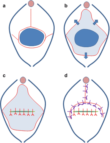
This incision is used for larger fistula. The incision starts at the posterior rim of the fistula and can be extended to the lateral walls of the vagina. The circumferential incision is then extended to the midline of the anterior vaginal wall allowing the development of two anterolateral flaps. This incision allows wide mobilization of the fistula tract and even of the urethra. The flaps can be used after the fistula closure to cover the fistula suture line (Fig. 11.2).


Fig. 11.2
(a) The large fistula is incised at the base. The incisions extend to the lateral vaginal wall and are extended in the midline of the anterior vaginal wall. (b) Dissection of the vaginal wall allows the formation of two anterolateral flaps, and a posterior flap and allows for wide mobilization of the fistula. (c) The fistula is closed in a tension-free way and the repair is watertight. (d) The flaps are used to cover the fistula suture line without creating an overlap between the suture lines
Ensure that the ureters are identified and eventually protected by placing ureteral catheters. If fistulas are located at the bladder neck, proximal urethra, or mid-urethra, make sure to maintain the urethral length as much as possible and think consider adding an autologous sling to prevent postoperative stress incontinence.
Closing of the Fistula
For the obstetric fistula, several consensus meetings have been held on the optimal closure of the fistula. Absorbable sutures 2/0 are recommended. Strong bites in the detrusor and serosal layers are necessary to close the fistula, but there is no need to close the urothelium separately. A single layer of separate sutures of 4–5 mm apart is sufficient to close the fistula. After closure, the bladder should be filled with blue dye to check for watertight closure. Eventually, supporting sutures can be used to stabilize the bladder neck. A Martius flap is optional in most cases [3].
Achieving and Maintaining Continence
Fistulas involving the bladder neck area or mid-urethra have a higher risk of persisting incontinence, despite a successful closure of the fistula. The urethral support mechanisms and the external urethral sphincter might be damaged as a consequence of the fistula, fibrotic process, or fistula repair. The larger the fistula, the more vaginal scarring and the smaller the bladder capacity, the higher the risk for postoperative incontinence. The surgical principles in these cases are the same as for other fistulas, but some additional measures can be taken. The urethral length should be maintained as much as possible. If the urethral length is less than 2.4 cm or if the urethral defect is more than 4 mm, an incontinence procedure should be added. If performed, postoperative incontinence can be reduced by approximately 50 %. Either a sling procedure can be added or a bladder neck suspension can be performed. For the sling procedure, only autologous material should be used. A sling can be created from fibromuscular tissue in the lateral vaginal wall or can be constructed out from the rectus or fascia lata. In the bladder neck suspension, non-absorbable sutures are used to suspend the bladder neck area in a fixed position [4].
Postoperative Care
Bladder drainage should be continued for 10–14 days, in most cases. In smaller fistulas that were easy to repair, shorter catheterization periods might be possible. When draining the bladder, silicone catheters are preferred since these have a larger internal diameter for the same external diameter and are less likely to be blocked by blot clots. A high fluid intake should be maintained to prevent clot formation. There is no need for antibiotics during the catheterization periods. Nursing staff should be trained and supervised adequately to ensure that catheters are not blocked, which could lead to a rupture of the fistula repair.
Abdominal Approach
Abdominal fistula repair can be done open, laparoscopically, or robotically. Extravesical and transvesical techniques have been described. The principles remain the same: identify the fistula, separate the bladder wall from the vaginal wall, and mobilize the fistula well enough to allow tension-free closure. Omentum can be used as interposition material.
Points of Interest
Most vesicovaginal fistulas can be managed by a vaginal approach, but abdominal, laparoscopic, or robotic repairs are also feasible.
Wide mobilization of the fistula to allow tension-free and watertight closure is of the utmost importance.
The timing of fistula surgery can be individualized, except for radiation fistula, where a longer waiting time (6–12 m) is advocated.
Bladder Augmentation
(5)
University of Lübeck, Lübeck, Germany
General Introduction
Many patients with small-capacity, high-pressure, or unstable or low compliant bladders will be managed with conservative measures. A small but significant minority of these patients will require surgical intervention, the therapeutic goals of which are to provide urinary storage while preserving renal function, continence, resistance to infection, and convenient voluntary and complete emptying.
Augmentation cystoplasty increases bladder capacity and decreases detrusor overactivity by enlarging the bladder with the addition of a bowel segment and possibly by disrupting the detrusor. Many different surgeons have carried out augmentation for many reasons, using many different types of tissue. The stomach, ileum, cecum, and ascending and sigmoid colon have all been used as tubular or detubularized, simple or complex segments.
The classic, augmentation enterocystoplasty has been performed in patients with neurogenic voiding dysfunction, but it has been shown to be effective for patients with neurogenic and non-neurogenic detrusor overactivity or patients with a reduced bladder compliance/capacity.
< div class='tao-gold-member'>
Only gold members can continue reading. Log In or Register to continue
Stay updated, free articles. Join our Telegram channel

Full access? Get Clinical Tree


