Fig. 8.1
Mesh fixation in TAPP with fibrin glue
Independently of this adhesive effect, it is advisable to take into account that some of these substances have been associated with a series of collateral effects which can be of benefit to the surgeon, these being either hemostasis actions favoring the closure of surgical wounds and line sealants of suture lines in anastomosis or favoring the handling of intestinal fistulas.
Tissue adhesives can be classified in various forms according to various parameters such as those shown in Tables 8.1 and 8.2.
Table 8.1
Classification of tissue adhesives based on its composition
Naturals | Fibrin (homologous) | Tissucol® o Tisseel® (Baxter, Westlake Village, CA, USA) |
Fibrin (autologous) | Vivostat® (Vivolution A/S, Birkerød, Denmark) Cryoseal® (Thermogenesis, Rancho Cordova, CA, USA) | |
Semisynthetics | Bovine albumin andglutaraldehyde | BioGlue® (Cryolife, Kennesaw, GA, USA) |
Synthetics | Cyanoacrylate | Histoacryl® (Braun, Aesculap AG, Tuttlingen, Germany) Glubran 2® (GEM Srl, Viareggio, Italy) Dermabond® (J&J, Somerville, NJ, USA) Indermil® (Covidien, Norwalk, CT, USA) |
Table 8.2
Classification of tissue adhesives based on their mechanism of action and secondary actions
Groups of adhesives/main composition | Primary mechanism of action | Properties derived from its primarymechanism of action |
|---|---|---|
Fibrin glue | Formation of a stable fibrin clot | Hemostatic, sealant, adhesive, healing |
Bovine albumin/glutaraldehyde | Bonding of tissues andnatural-synthetic structures | Adhesive (sealant, hemostat) |
Cyanoacrylate | Bonding of tissues andnatural-synthetic structures | Adhesive (sealant, hemostat) |
Using the Liechtenstein method on 21 patients with inguinal hernias, Farouk et al. in 1996 [4] described the repair of a series of hernias using butyl-2-cyanoacrylate adhesive. The study received some critical reviews related to the toxicity of this adhesive associated with the production of local heat and a consequent lesion of tissues and nervous structures. In 1997, Chevrel and Rath [5] described for the first time the use of fibrin glue (FG), a biological glue previously used for the closure of surgical wounds in the fixation of meshes during abdominal hernia surgery.
Various studies have analyzed the advantages and limitations of the different commercial types of FG: Tissucol® (Baxter, Vienna, Austria), Quixil® (Omrix Biopharmaceuticals, Ethicon Inc. Somerville, NJ, USA), and Vivostat® (Vivolution A/S, Birkerød, Denmark), with the aim of achieving an optimal integration of FG in the surrounding tissues. First of all, the mechanism effect of Tissucol® and Quixil® is based on the reproduction of the final steps of coagulation cascade, thanks to its simultaneous application on its two components: (1) concentrate of human fibrinogen and factor XIII lyophilization, which is reconstituted with aprotinin (antifibrinolytic) and (2) thrombin, reconstituted with calcium chloride. The difference between both lies in thrombin of bovine origin in the first and of human origin in the second.
For its part, Vivostat® is an autologous FG created from the extraction of 120 ml of a patient’s blood mixed with sodium citrate. Autologous FG provides increased safety compared to non-autologous FG. The use of the patient’s own blood eliminates the risk of the introduction into the organism of infectious material of human or animal origin, as well as hypersensitivity reactions to human and bovine proteins. However, for the time being, there has been no evidence of the transmission of viral hepatitis or HIV infection by this means. In fact, Tissucol® uses plasma obtained in official European plasmapheresis centers and is submitted to a screening process for antigens and antibodies beforehand through thermal inactivation.
The results of the use of autologous and non-autologous FG were compared in a case–control study. It focused on surgical meshes Vypro II® (Ethicon Inc., Norderstedt, Germany) in a group of 20 patients with autologous FG (Vivostat®, Vivolution A/S, Birkerød, Denmark). The results were compared with a group of 20 patients in which the mesh was fixed with non-autologous FG (Tissucol®, Baxter, Vienna, Austria). The authors highlighted that the level of performance of both products was similar. As a matter of fact, no differences were found in the amount of complications at day 7 and at 6 months, although the cost of autologous FG was higher [6, 7].
Traumatic Fixation Methods
For mechanical traumatic fixation, we can opt for long-life absorbable staples (reabsorption in 1 year) made of polyglycolic and polylactic acid, which maintain tensile strength for up to 3–5 months (Fig. 8.2). Alternatively, we can opt for nonabsorbable staples (Fig. 8.3), usually made of titanium (an inert material, biocompatible and hypoallergenic), which provide a great resistance to traction, making them suitable when attempting to obtain a good grip on the tissues, which guarantees anchorage and firm fastening. They are presented in different formats by different companies (Table 8.3). It is convenient to remember that at present, there are plastic nonabsorbable fixation methods on the market.
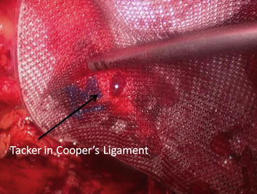
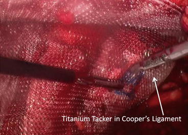
Fig. 8.2
Absorbable tacker in Cooper’s ligament
Fig. 8.3
Titanium tacker in Cooper’s ligament
Table 8.3
Devices for mechanical traumatic fixation
Staplers | Metallics | Absorbables | Plastics |
|---|---|---|---|
Helicoidals | ProTack® (titanium) 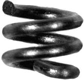 | AbsorbaTack® (massif) 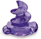 | |
SorbaFix® (hollow)  | permafix® (hollow)  | ||
U-shaped | EMS® Stapler®  | Securestrap®  | |
Circulars | SALUTE® 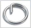 | ||
Others | PermaSorb®  | ||
I-Clip®  |
When proceeding with the fixation of the mesh using staples or tackers, and with the aim of minimizing the risk of lesions and chronic pain, which is the main inconvenience connected to their use, it will be vital to know with accuracy the anatomy of the area and the location of inguinal nerves. Staples or tackers should be avoided in the “triangle of pain” and in the “triangle of doom” as well as in the area of exposure of nerves by leaving the fascia protector of the nerve intact and minimizing the use of electrocautery (Fig. 8.4). It is well known that the traumatic anchorage of the mesh to the pubis will invariably determine postoperative pain in a high percentage of cases, although many laparoscopic surgeons continue to carry this out. It is fundamental to avoid these “hot spots” for the development of pain and lesions, as well as reduce the number of tackers used in each patient (maximum of 4), in order to improve results in the short and long term.

Fig. 8.4
Traumatic fixation must respect the triangle of doom and the triangle of pain
Fixation Versus Non-fixation
With the aim of minimizing the risk of chronic pain comes the idea of eliminating traumatic fixation of the meshes, as this type of system is associated with possible nerve entrapment causing acute long-term pain, as well as a higher risk of vascular and secondary visceral lesions. As a result of this, different authors started to consider not fixing the mesh as an alternative, analyzing whether this increased the recurrence rate. This showed a recurrence rate of between 0.7 and 0.5 % in those controlled studies which compared fixation with staples to non-fixation, respectively, and between 0.6 and 0.4 % in those which compared fixation with staples compared to fixation with fibrin. With regard to chronic pain, the different studies published highlighted a significant reduction in chronic pain in those patients which were operated on using staple fixation, as opposed to those in which the mesh was not fixed. Likewise, there was a reduction of pain in those patients which were operated on using fibrin fixation as opposed to fixation with staples. However, controlled studies do not currently exist which compare non-fixation to fixation with fibrin. There are also few cases which compare fixation with absorbable staples; therefore, it is not possible to reach a conclusion.
As a result of this, “non-fixation” of the mesh is proposed as a cost-effective alternative, since in the different published series, it is not seen as increasing the recurrence rate, and it is also associated with a lower cost, less operating time, and a shorter stay in hospital. It is due to these points that a policy of non-fixation is recommended, but in a selective manner, depending on the type of hernia. As a result of this, non-fixation of the mesh could be considered in the case of types L I-II and M I-II hernias, while in cases of direct and large hernias (MIII and LIII), fixation using fibrin glue for the fixation of the mesh is recommended, minimizing the risk of acute and chronic pain, as opposed to the use of staples.
Related posts:
 Prostheses in Laparoscopic Inguinal Hernia Repair
 Emergency Laparoscopic Surgery of the Abdominal Wall
 Basic Concepts in Laparoscopic Hernia Repair
 Fixation of Prostheses in Laparoscopic Ventral Hernia Repair
 Current Advances and New Frontiers in Laparoscopic Hernia Repair
 Laparoscopic Approach in Other Hernias: Subcostal, Xiphoid, Lumbar, Suprapubic, Parastomal, and Spigelian
Stay updated, free articles. Join our Telegram channel

Full access? Get Clinical Tree


