Congenital
Osseuos
Benign
Benign
Developmental cysts
Giant cell tumor
Rectal duplication
Osteoblastoma
Anterior sacral meningocele
Aneurysmal bone cyst
Adrenal rest tumor
Malignant
Malignant
Osteogenic sarcoma
Chordoma
Ewing sarcoma
Teratocarcinoma
Myeloma
Neurogenic
Chondrosarcoma
Benign
Miscellaneous
Neurofibroma
Benign
Schwanomma
Lipoma
Ganglioneuroma
Fibroma
Malignant
Leiomyoma
Neuroblastoma
Hemangioma
Ganglioneuroblastoma
Endothelioma
Ependymoma
Desmoid
Malignant peripheral nerve sheath tumors
Malignant
Liposarcoma
Fibrosarcoma/malignant fibrous histiosarcoma
Leiomyosarcoma
Metastatic carcinoma
Other
54.2 Anatomy
A complete understanding of the anatomy of the presacral space is essential in the management of these tumors. The presacral space is a potential space bounded superiorly by the peritoneal reflection and inferiorly by the retrosacral fascia (which extends from the S4 vertebra to the rectum a few centimeters cephalad to the anorectal junction). The mesorectum is the anterior border and the posterior border is composed of the presacral fascia. Laterally, the space is defined by the ureters, the iliac vasculature, and sacral nerve roots (Fig. 54.1).
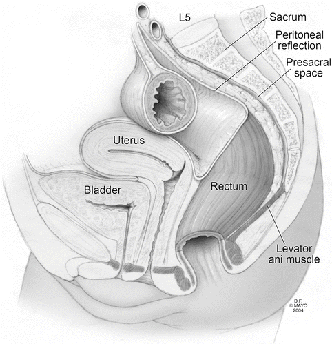
Fig. 54.1
The presacral space (By permission of Mayo Foundation for Medical Education and Research. All rights reserved)
Presacral tumors may arise from or locally invade pelvic structures (Fig. 54.2). In malignant cases, these structures may need to be sacrificed at the time of resection if an oncologically appropriate operation is done. Due to the high risk of injury and resection of local-regional structures, it is important to counsel patients on the potential for anorectal, sexual, or physical debility following surgery. Loss of bilateral S3 nerve roots will result in incontinence and mandates an end colostomy. If all sacral nerve roots on either side are sacrificed, but one side is preserved, normal function will be maintained [5]. High ligation of the inferior mesenteric artery, or during mobilization of the rectum near the sacral promontory, may result in injury to the hypogastric nerves resulting in retrograde ejaculation and/or bladder dysfunction [6]. Injury to the Nervi erigentes (parasympathetic fibers from S2 to S4), which course anteriorly in the lateral stalks, may result in erectile dysfunction. The pudendal nerve (S2–S4) courses inferiorly to the perineum—a sensory branch carries fibers to the skin of the penis and glans and a motor branch innervates the external anal sphincter. A unilateral pudendal nerve injury does not generally result in incontinence because there is cross innervation of the fibers between the right and left pudendal nerves at the level of the spinal cord [7].
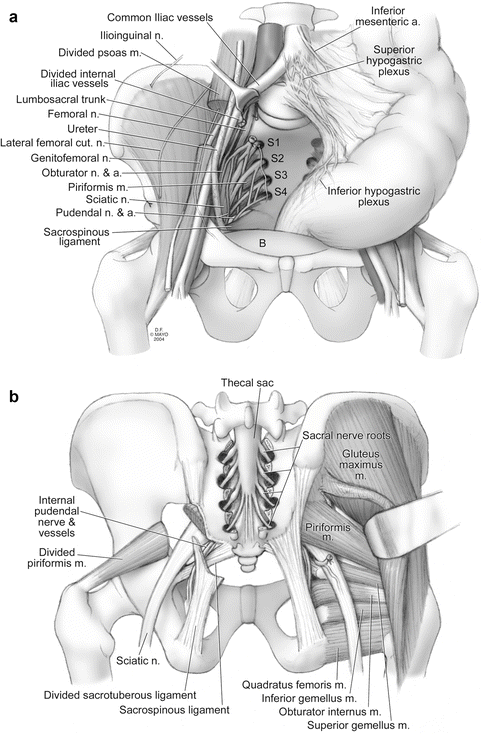
Fig. 54.2
Neurovascular relationships of the pelvis. (a) Anterior view. (b) Posterior view (By permission of Mayo Foundation for Medical Education and Research. All rights reserved)
If adequate resection necessitates sacrectomy, our practice utilizes the skills of an oncologic orthopedic and spine surgeon to assist. Familiarity of the bony pelvis, spinal nerve roots, and pelvic ligaments and musculature is imperative for safe conduct of the operation. The majority of the sacrum can be safely removed with stability maintained in non-irradiated patients if more than half of the S1 vertebral body is preserved.
54.3 Diagnosis
Presacral tumors are often found incidentally and diagnosis requires a high degree of clinical acumen. Symptoms, when present, are often vague and non-specific [8]. Those patients that complain of pain (pelvic, perineal) more often present with malignant tumors, and those with neurologic dysfunction (urinary/fecal incontinence, sexual dysfunction), also often have more advanced tumors [1, 9]. Some patients complain of perineal drainage or posterior midline dimpling—leading to a misdiagnosis of pilonidal disease or perianal fistula [10].
A comprehensive neurologic examination must be completed to define any deficits and to document function preoperatively. In a series from the Mayo Clinic, 97 % of presacral tumors could be palpated on digital rectal examination [1]. Digital rectal examination aides in defining the proximal extent of the lesion, and tumor fixation to pelvic structures. Endoscopic evaluation should be completed to evaluate for penetration into the rectal lumen or other synchronous colorectal lesions.
Evaluation of presacral lesions is greatly enhanced by computed tomography (CT) and/or magnetic resonance imaging (MRI) with interpretation by a radiologist specializing in musculoskeletal disease. These imaging modalities are complimentary and aid in operative planning. MRI has superior soft-tissue contrast resolution which may provide improved determination of the anatomic extent of the tumor [11]. MRI is also more sensitive than CT in spinal imaging—demonstrating cord abnormalities (meningocele, nerve root or thecal sac involvement) [12]. CT has a superior ability to evaluate cortical bone destruction. Imaging will determine more accurately than physical exam whether a lesion is cystic, solid, or heterogeneous and whether adjacent pelvic structures are involved.
Some patients who present with a presacral tumor will have a rare congenital disorder called Currarino Syndrome. Currarino Syndrome is an autosomal dominant disorder characterized by: (1) sacral anomalies, (2) presacral masses (such as an anterior sacral meningocele or teratoma, sometimes more than one), and (3) anorectal malformations [13, 14] (Fig. 54.3). Taking a detailed family history will often reveal a consistent autosomal dominant pattern of these anomalies.
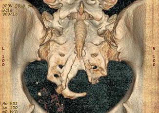
Fig. 54.3
Three-dimensional CT reconstruction of the posterior sacrum demonstrating scimitar anomaly, commonly seen in Currarino syndrome (By permission of Mayo Foundation for Medical Education and Research. All rights reserved)
Preoperative determination of benign versus malignant status of presacral tumors is critical to make decisions on use of adjuvant therapy and plan lines of surgical resection. Advancements in modern imaging techniques have improved the ability to identify malignant lesions without a biopsy. Some authors, in fact, believe that biopsy of presacral lesions is contraindicated and unnecessary in tumors deemed resectable [1, 9, 15, 16]. In a recent review by the Mayo Clinic, preoperative biopsy of solid or heterogeneous tumors demonstrated a high concordance with post-operative pathology compared to imaging alone (91 % vs. 36 %) with a low complication rate. Moreover, almost half of patients could not be given a definitive diagnosis based on imaging alone and of those over one-third were found to be malignant tumors [17]. Simple cystic lesions are usually not biopsied as management is generally not altered by biopsy results. Patients with malignant tumors responsive to neoadjuvant chemoradiation (Ewing sarcoma, osteogenic sarcoma, neurofibrosarcoma, etc.) will have tissue confirmation of their disease prior to initiating therapy. Additionally, biopsy results will provide information regarding the need for wide en-bloc oncologic resection verses a potentially limited, nerve- or function-sparing intralesional resection. In our practice, all solid or heterogeneous tumors undergo pre-operative percutaneous (most often either trans-sacral or trans-perineal) biopsy and no tumor is biopsied trans-rectally or trans-vaginally. Route of biopsy should be planned to be within the field of proposed resection so that the tract may be excised if necessary (Fig. 54.4).
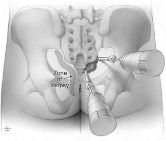
Fig. 54.4
Zones of suggested CT guided biopsy, within the field of proposed resection (By permission of Mayo Foundation for Medical Education and Research. All rights reserved)
54.4 Surgical Treatment
The primary treatment modality of presacral tumors is operative removal. Reasons for an aggressive approach are obvious for malignant tumors. For benign lesions, there exists a known risk of malignant transformation with certain tumors [18]. Some lesions may obstruct the vaginal canal and lead to complicated vaginal delivery [19]. An untreated anterior sacral meningocele may become infected resulting in potentially life-threatening meningitis.
Related posts:
 Radical Cystectomy: Robotic, Laparoscopic, Open and Partial
 Diagnostic and Endoscopic Management of Bladder Tumors
 Considerations for Management of Hereditary Rectal Cancer and Desmoid Tumors
 Training Surgeons for Rectal Cancer Surgery: Clinical and Simulation
 Radical Hysterectomy in Cervical Cancer
 Imaging of Gynecological Cancers
Stay updated, free articles. Join our Telegram channel

Full access? Get Clinical Tree


