Fig. 9.1
Image of a female patient with an obstructing distal stone in the common ureter of her ureterosigmoidostomy. She was treated successfully with antegrade ureteroscopy
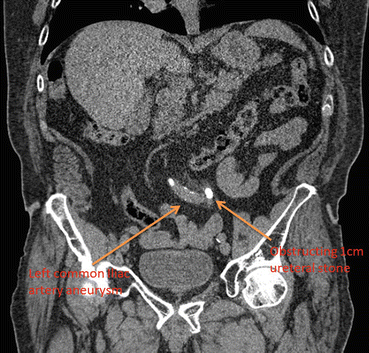
Fig. 9.2
Seventy-eight year old male with calcified common iliac artery aneurysm and 1 cm obstructing ureteral calculi
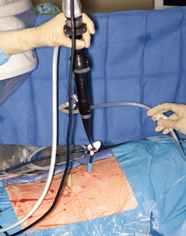
Fig. 9.3
Antergrade ureteroscopy performed with the two access sheath technique. This particular patient had a large lower and upper pole stone burden in addition to an impacted ureteral stone. Thus, two renal access sheaths are in place, one for the lower pole stones and one to access the upper pole and ureteral stone. A ureteral access sheath has been advanced through the upper pole access
Preoperatively patients are counseled in the outpatient setting regarding the risks of antegrade URS and the typical convalescence. If a new percutaneous tract is established, overnight observation is recommended to assess for any significant renal bleeding. If an established tract is utilized (i.e. patient already has indwelling nephrostomy tube), then the surgery may be performed as an outpatient procedure. All patients have a urinalysis with culture obtained as well as complete blood count and basic metabolic panel. All positive urine cultures should be treated prior to the procedure with culture-specific antibiotics. We recommend recent computed tomography (CT) imaging within the last 30 days to confirm stone size and location. Three dimensional CT imaging also provides information on surrounding organs and renal anatomy allowing for appropriate preoperative planning for the percutaneous renal access. If a new percutaneous tract into the kidney is created, a type and cross is obtained and patients are consented regarding the risk of blood transfusion.
Equipment
To perform percutaneous antegrade ureteroscopy the surgeon will require standard equipment for both percutaneous nephrolithotomy and for ureteroscopy. We recommend a rigid nephroscope, flexible nephroscope, and flexible ureteroscopy. Percutaneous access and establishing a percutaneous access tract using a renal access sheath should be performed as previously described [18]. Once a percutaneous renal access sheath is in place there should be a working wire in the ureter. A ureteral access sheath (13 or 14Fr outside diameter) can be placed through the 30Fr renal access sheath to the level of the ureteral stone. The ureteral access sheath facilitates stone removal from the ureter in an efficient manner (Fig. 9.3). A laser lithotripter, as well as stone basket for retrieval of fragments, is recommended. An ultrasonic suction device can also be helpful to remove stone debris or blood clots that collect in the renal pelvis at the time of the procedure. A ureteral stent and/or nephrostomy tube is used at the conclusion of the case.
Patient Positioning
We prefer placement of the patient in the prone position on a C-arm capable bed with careful attention paid to pad all pressure points. If we anticipate need for retrograde access to the ureter at time of antegrade ureteroscopy, then a prone split leg position is utilized. Stuurman and colleagues placed patients in both the modified supine position with elevation of the treated side and the modified Valdivia position, in which the patient is placed in a split leg position with the treated side raised as well in order to treat patients with urinary diversions [17].
Description of Technique
An understanding of renal and ureteral anatomy is vital when attempting antegrade ureteroscopy in particular when retrograde access has failed due to distal ureteral pathology. Our current practice utilizes triangulation technique for upper-pole percutaneous access as previously described [18]. Upper pole access is preferred as it allows for a direct course into the proximal ureter. Once access is established, a 0.038-in. hydrophilic nitinol glidewire is advanced down the ureter to the level of the stone. The wire is then exchanged for a stiff workable wire and the tract is then dilated using an 8/10Fr dilating catheter and a second stiff safety wire is placed through the 10Fr dilator. The working stiff wire is then used to advance the balloon dilator into the collecting system. The tract is balloon dilated and a 30Fr renal access sheath is then advanced into the kidney. Rigid nephroscopy is then performed to confirm appropriate sheath placement. An ultrasonic lithotripter with suction capabilities is often helpful to remove any blood clot or debris that may be present in the renal pelvis. Flexible nephroscopy is then performed to inspect the entire kidney and proximal ureter. If the flexible nephroscope cannot be easily advanced to the ureteral stone then a flexible ureteroscope will be necessary to complete stone removal. In order to facilitate easy access to and from the stone with the ureteroscope it is often beneficial to place a ureteral access sheath through the renal access sheath. Placement of the ureteral access sheath to the level of the stone is performed using a 12/14 or 11/13Fr sheath advanced with fluoroscopic guidance over the stiff working wire.
The ureteroscope is then easily advanced through the ureteral sheath to the stone. Once the stone is visualized laser lithotripsy is performed utilizing the holmium laser with settings appropriate for stone fragmentation (our preference is 8 Hz and 0.8 J). Stone fragments are then removed utilizing basket extraction. After all sizeable fragments have been removed, a guidewire is advanced down the length of the ureter and URS is performed for a full inspection of the ureter to the interior of the bladder. The ureteral access sheath is removed and the kidney is inspected one last time with the rigid and flexible nephroscope to remove any debris which may have migrated proximally. Next a double-J ureteral stent is deployed under fluoroscopic guidance. We generally leave the stent indwelling for 2–4 weeks depending on the degree of stone impaction and ureteral condition.
While our practice utilizes the “double sheath” technique for antegrade URS, a single ureteral access sheath-only technique can be utilized as well. Winter and colleagues describe a “no dilation” or “minimal dilation” approach with only the 12/14Fr access sheath passed antegrade over a guidewire which has already been passed down the ureter [19]. We prefer to have a renal access sheath in place to remove proximally displaced stones and concomitant renal stones which cannot be done with the single sheath technique.
If upper pole access cannot be obtained due to patient anatomy, a mid-pole or even lower pole access can be utilized. However, if the angle for the calyx to the ureter is too great then a ureteral access sheath cannot be used. In such cases, the ureteroscope is advanced down the ureter to the level of the stone visually. The stone material can then dusted it to very fine debris to be passed, with larger fragments extracted by basket. Such a technique can be very time consuming and tedious and is only recommended if all previously described options have been deemed inappropriate.
Related posts:
 Radiation Exposure to the Patient and the Urologist
Shock Wave Lithotripsy for the Treatment of Ureteral Stones
Selecting the Appropriate Treatment Modality for Ureteral Calculi
Radiation Exposure to the Patient and the Urologist
Shock Wave Lithotripsy for the Treatment of Ureteral Stones
Selecting the Appropriate Treatment Modality for Ureteral Calculi
 Semirigid Ureteroscopy for Ureteral Calculi
 Tips and Tricks in the Treatment of Ureteral Stones
 Adjunctive Equipment for Ureteral Stone Management
Stay updated, free articles. Join our Telegram channel

Full access? Get Clinical Tree


