Primary tumour (T)
TX
Primary tumour cannot be assessed
T0
No evidence of primary tumour
Tis
Carcinoma in situ
Ta
Non-invasive verrucous carcinoma
T1
Tumour invades subepithelial connective tissue
T1a
Tumour invades subepithelial connective tissue without lymph vascular invasion and is not poorly differentiated
T1b
Tumour invades subepithelial connective tissue with lymph vascular invasion and is poorly differentiated
T2
Tumour invades corpus spongiosum or cavernosum
T3
Tumour invades urethra
T4
Tumour invades other adjacent structures
Regional lymph nodes (N)
Clinical stage (palpation + imaging)
cNX
Regional lymph nodes cannot be assessed
cN0
No palpable or visibly enlarged inguinal LN
cN1
Palpable mobile unilateral inguinal LN
cN2
Palpable mobile multiple or bilateral inguinal LN
cN3
Palpable fixed inguinal LN mass or pelvic lymphadenopathy unilateral or bilateral
Pathologic stage (biopsy or surgical excision)
pTNX
Regional lymph nodes cannot be assessed
pTN0
No regional lymph node metastasis
pTN1
Metastasis in a single inguinal lymph node
pTN2
Metastasis in a multiple or bilateral inguinal lymph nodes
pTN3
Extranodal extension of lymph node metastasis or pelvic lymph node(s) unilateral or bilateral
Distant metastasis
M0
No distant metastasis
M1
Distant metastasis (LN metastasis outside of the true pelvis in addition to visceral or bone sites)
Anatomic stage/prognostic groups
Stage 0
TisN0M0 or TaN0M0
Stage I
T1aN0M0
Stage II
T1bN0M0 or T2N0M0 or T3N0M0
Stage IIIa
T1–3N1M0
Stage IIIb
T1–3N2M0
Stage IV
T4, any N, M0 or any T, N3, any M or any T, any N, M1
24.5 Diagnosis of the Primary Lesion
The patient presents generally after several weeks or months due to negligence or denial. Sometimes the lesion remains unnoticed being hidden by the prepuce in 25–75 % cases. Overall 30 % of penile cancer are diagnosed at advanced stage [11]. The presenting complaints are consistent with a painless non-healing ulcer. This can be associated with itching, burning sensation and oozing. Sometimes there is intermittent bleeding (either spontaneously or after a scratch), foul-smelling discharge and even dysuria in advanced cases. The diagnostic approach is initiated by a physical examination to assess the primary tumour and evaluate inguinal metastatic lymph nodes when present. A penile lesion that does not resolve after 2–3 weeks of careful observation and skin care requires biopsy. Preferably excision biopsies should include enough tissue to determine the depth of invasion for accurate staging and subsequent planning of treatment. An incisional biopsy is recommended for large lesions [12].
At presentation, squamous cell carcinoma is found in the glans in 48 % of cases, in the prepuce in 21 %, in both glans and prepuce in 9 %, on the coronal sulcus in 6 % and in the shaft in <2 % [13].
In the past about 50 % of palpable inguinal LN (cN1–cN2) at presentation were considered to be inflammatory [13], and it was common practice to first treat the patient with fluoroquinolones for 4–6 weeks. Only after that period were persistent LNs considered as metastatic and managed accordingly. Nowadays expert panels consider this old dogma no longer valid because metastatic disease is highly likely to be present in these patients. It is now recommended to proceed to fine needle aspiration cytology (FNAC) in all patients without delay, even in those with non-palpable LN where US guidance should be used [14, 15]. Furthermore, ILND should be considered for all diseases higher than pT1G2 even in N0 patients [14]. Histopathology of the primary lesion is of paramount importance in predicting the risk of locoregional extension: this is increased by the presence of vascular, neural or lymphatic invasion, a deep or high-grade tumour or a sarcomatoid variant which has a 90 % risk of nodal extension. A study has shown that almost all patients with tumour depth of invasion greater than 6 μm and all patients with vascular invasion developed cancer progression after a mean follow-up of 28 months [16]. Conversely Tis, pTa and verrucous carcinomas are never associated with nodal disease [17]. Buschke-Löwenstein lesion or giant condyloma acumunata is a variant of verrucous carcinoma, a well-differentiated form of SCC representing about 5 % of penile SCC. It does not metastasize, but it is clinically important due to its ability to spread by local extension and eventually to destroy part or all the penile tissues if left untreated [18].
MRI imaging after intracavernosal contrast has shown great potential in local staging, accurately predicting corpora cavernosa invasion compared to histopathology, and was proven to be a reliable tool in selecting patients for partial penectomy [19].
Tomoscintigraphy, a combination of CT scan with scintigraphy after technetium 99, also called SPECT (single-photon emission computed tomography) is being evaluated for early sentinel lymph node detection [20].
Note: A difficult differential diagnosis may exist with an extramammary Paget’s disease of the penis. This lesion presents as a penoscrotal erythematous lesion progressing for several months and resistant to antifungal drugs. It is in fact an intraepithelial adenocarcinoma often associated with a regional malignancy (prostate, bladder, colon, rectum) and readily diagnosed by biopsy. Isolated Paget’s disease of the penis is extremely rare [21].
24.6 Disease Extension Imaging
Despite having low sensitivity for detecting pelvic or para-aortic LN metastases of a penile cancer, CT scan remains the standard investigation because MRI does not perform any better [22]. Nonetheless CT is only indicated if inguinal LNs are positive, because there is no pelvic or aortic LNs invasion without initial involvement of the inguinal LNs [23, 24].
PET-TDM using 18F-FDG has been shown to have good sensitivity in N1 disease or higher and excellent specificity with an overall diagnostic accuracy of 89 % in detecting nodal and distant metastases of a penile cancer. It is recommended as a second imaging modality if inguinal LNs are clinically palpable when a suspicion of pelvic or distant disease persists after an inconclusive or negative CT scan [25, 26].
24.7 Treatment
24.7.1 Tis and Ta
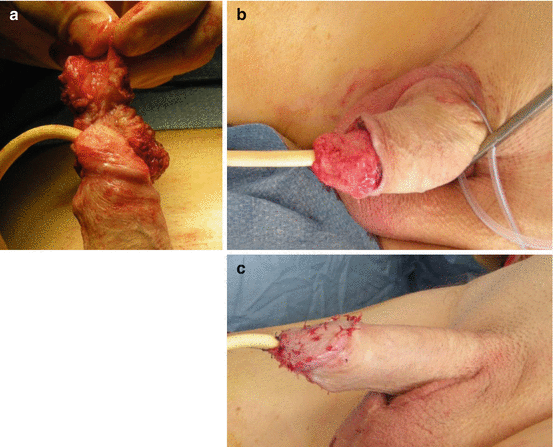
If isolated small preputial lesion, a circumcision can be sufficient. Systematic circumcision is recommended even in balanic localization. An associated partial glansectomy remains a possibility for relatively large lesion provided there is a 1–2 mm healthy margin. Mohs technique (serial progressive excision + histopathology) is preferable in such cases in order to ensure negative margins with minimal excision [27]. CO2 and Nd-Yag (neodymium-doped yttrium aluminium garnet) lasers are also suggested as organ-sparing tools [28]. Other conservative measures are cytotoxic cream (5-fluorouracil, imiquimod 5 %) and dynamic phototherapy. Surveillance is essential after these conservative procedures with reassessment every 3 months for 2 years. Complete removal of a non-invasive verrucous carcinoma can be achieved by thorough “shaving” of the lesion with optimal organ preservation (Fig. 24.1a–c).


Fig. 24.1
(a–c) Shaving of a non-invasive verrucous carcinoma developed onto the glans. The growth was removed with a complete organ preservation. Figure (c) shows a split-thickness skin graft harvested from the thigh using a 0.5 mm dermatome (Courtesy N. Morel-Journel, Urology, CHU Lyon, France)
24.7.2 T1
Same, but with a 3 mm margin. CO2 and Nd-Yag laser can be applied. Beware of the 32 % recurrence rate following organ preservation procedures [29].
24.7.3 T2
Involving solely the glans: partial glansectomy if the lesion occupies less than 50 % of the glans, otherwise total glansectomy.
Extending proximal to the glans: partial penectomy is advocated provided a 2–cm negative margin can be obtained and a minimum 3 cm penile length can be preserved to allow the patient to urinate without permanently soaking his scrotum. Otherwise, it is advised to proceed to a total penectomy and perineal urostomy. A total amputation can also be performed if the patient is not compliant with regular follow-up.
24.7.4 Management of Lymph Nodes
Ultrasonography and CT scan may be useful in the presence of an obese patient with LNs that are not palpable but in whom there is a strong clinical and pathological suspicion of nodal extension. If enlarged nodes are found by US, they should be needle-biopsied. If biopsy is positive, LN dissection is indicated. If biopsy is negative, tomoscintigraphy should be performed to exclude a sentinel node. If this is found, it should be removed along with the palpable LNs.
< div class='tao-gold-member'>
Only gold members can continue reading. Log In or Register to continue
Stay updated, free articles. Join our Telegram channel

Full access? Get Clinical Tree


