Fig. 2.1
Lumbosacral dematomes, cutaneous nerves, and reflexes. (Panicker JN et al. Lancet Neurol 2015)
Urodynamic investigation, with simultaneous fluoroscopic monitoring (i.e., video-urodynamics), is essential to assess detrusor and bladder outlet function, and it is crucial for clinical decision making. Generally accepted risk factors jeopardising the upper urinary tract are high detrusor pressure during storage phase due to low-compliance bladder and/or detrusor overactivity combined with detrusor sphincter dyssynergia, and urodynamic investigations are needed to identify these conditions.
Urethrocystoscopy (combined with bladder washing cytology if appropriate) is used if indicated to detect urethral and bladder pathologies, such as urethral stricture, urethral or bladder stones and bladder tumours, including carcinoma in situ.
Serum creatinine, cystatin c and corresponding estimations yield a reasonable estimation of renal function with minimal cost and inconvenience. Creatinine clearance provides a more accurate assessment but involves a 24-h urine collection to measure creatinine excretion. This may result in underestimation of renal function if the urine collection is incomplete. The most accurate measurement is isotopic glomerular filtration rate, especially when renal function is poor or with alterations of muscle mass (as is common in patients with neurological disease).
Clinical Practice
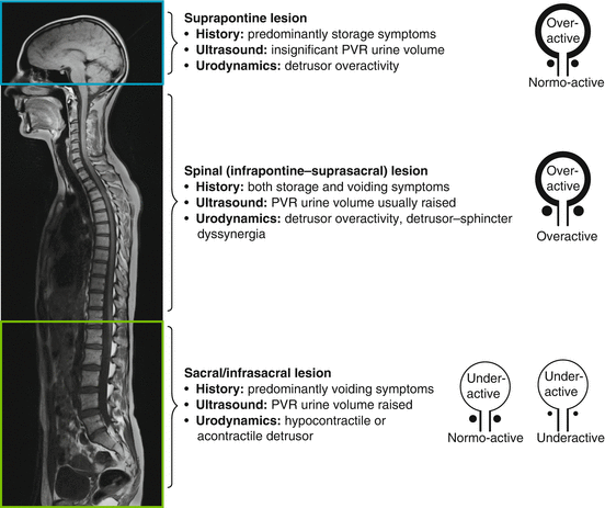
A very simple classification system for use in daily clinical practice is provided in (Fig. 2.2

). Based on the dysfunction pattern, the appropriate therapeutic strategy to preserve both upper and lower urinary tract function, and to achieve or maintain urinary continence, is determined.

Fig. 2.2
Patterns of lower urinary tract dysfunction following neurological disease. (Panicker JN et al. Lancet Neurol 2015)
In patients with detrusor overactivity, the therapeutic concept is to convert the overactive detrusor into a normoactive or underactive one. Commonly, antimuscarinics are the pharmacological treatment of choice, but they have limited effectiveness and many patients discontinue their use due to adverse events. The beta-3 adrenergic agonist mirabegron has recently been introduced as an alternative to antimuscarinics for treatment of idiopathic overactive bladder, but research into its application in neurogenic LUTD has not been reported thus far. For refractory neurogenic detrusor overactivity, intradetrusor onabotulinumtoxinA injections are a highly effective, minimally invasive and generally well-tolerated treatment that improves health-related quality of life. In the case of failed onabotulinumtoxinA treatment, augmentation cystoplasty is a well-established treatment option but requires abdominal surgery with interposition of an intestinal segment (usually ileum) into the bladder and/or partial replacement of bladder by an intestinal substitute. In highly selected patients, cystectomy with continent or incontinent urinary diversion becomes necessary as a salvage procedure.
In patients with underactive/acontractile detrusor and/or with detrusor sphincter dyssynergia, intermittent self-catheterisation is recommended to assist bladder emptying. Passive voiding by abdominal straining (Valsalva manoeuvre) or, particularly, by suprapubic downwards compression of the lower abdomen (Credé manoeuvre) is not recommended as it creates high, unphysiological intravesical pressure which puts the upper urinary tract at risk and causes compression of the urethra, i.e. a functional obstruction that leads to inefficient emptying. Nevertheless, some patients are not able and/or not willing to perform intermittent self-catheterisation, and therefore, an indwelling transurethral or suprapubic catheter is potentially the only alternative.
In the case of stress urinary incontinence due to low bladder outlet resistance, electrical stimulation of the pelvic floor can help to restore urinary continence in patients with incomplete lesions. In some neurological patients, the implantation of a sub-urethral sling or an artificial urinary sphincter may become necessary. However, it needs to be considered that artificial urinary sphincters generally do not continue working indefinitely and may need to be replaced with increased risks for revision surgery.
Regular follow-up is essential since neurogenic LUTD is often unstable and symptoms might vary considerably even within a relatively short period. The EAU Guidelines on Neuro-Urology provide clear-cut grade A recommendations that any significant clinical changes should instigate further specialised investigation and that in high-risk patients, the upper urinary tract should be assessed at least every 6 months, physical examination and urinalysis should take place every year, and urodynamics should be done at regular intervals. However, there is a complete lack of high-evidence level studies on which to base such recommendations. Thus, follow-up of the neuro-urological patient is more eminence- than evidence-based. There is no uniform follow-up, and a rather individualised, patient-tailored approach aiming to achieve an optimal quality of life and to protect the upper urinary tract is needed for this special population.
Spina Bifida
Background
Although spina bifida is one of the most common birth defects of the spine, the exact mechanisms resulting in closure or a dysraphic state are yet to be elucidated. Nevertheless, the aetiology of closure defects of the neural tube is supposed to be multifactorial, including both genetic and environmental factors. The deficit of folic acid in the early pregnancy period is a major risk; the role of folic acid in the prevention of neural tube defects has been established since the early 1990s. Maternal ingestion of 400 μg of folic acid per day in all women of child-bearing age can reduce the incidence of spina bifida by 50 %. In the United Kingdom and Ireland, the yearly prevalence of neural tube defects declined, predating any periconceptional folic acid supplementation policy initiatives from 4.5 per 1000 births in 1980 to 1–1.5 per 1000 in the 1990s.
In spina bifida patients, the neurological findings can vary from the most discrete deficit to a complete paraplegia potentially involving all levels of the spinal column. Most spinal defects occur at the lumbar spine with the sacral, thoracic and cervical areas, in decreasing order of frequency, less affected. Associated Arnold-Chiari malformation is seen in more than 80 % of spina bifida children mostly requiring ventriculoperitoneal shunt placement. Whilst the likelihood of bladder, bowel and pelvic floor dysfunction depends on the severity of the lesion, the pattern of the dysfunction is aligned to the localisation.
Neurogenic LUTD in children with spina bifida can lead to secondary upper urinary tract deterioration and often causes chronic urinary incontinence. The preservation of renal function is the primary goal in the neuro-urological management of spina bifida patients, but considering the impact on quality of life, efforts to promote urinary continence have become more and more important. Continence is associated with better self-concept, and incontinent girls are particularly at high risk for poor self-esteem. Urinary incontinence is a stress factor for these patients, and even a slight improvement in urinary continence means to these patients additional independence. Thus, reducing the frequency of incontinence or, even better, the achievement of continence is an important goal of urological medical care.
According to Verhoef et al., urinary and faecal incontinence is common in young adults with spina bifida (61 % and 34 %, respectively) regardless of bladder and bowel management they used. The majority of urinary and faecal incontinent patients perceived this as a problem (70 % and 77 %, respectively). Moreover, the authors of this study found that patients with a level of lesion at L5 or above were far more likely to be urinary and faecal incontinent than those with a level of lesion of S1 or below. However, patients with a level of lesion at L5 and above were also more likely to perceive urinary incontinence as a problem. It is well known that urinary incontinence in these patients has an underlying component of detrusor overactivity and/or poor bladder compliance, which is more frequent in patients with intact or at least partially intact sacral reflexes, which are mostly present in lesions at L5 and above (suprasacral lesion). Remarkably, whilst in utero closure of spina bifida decreased rates of ventriculoperitoneal shunting and improved motor function, it seems not to be associated with a relevant improvement in lower urinary tract function compared to repair after birth.
Points of Interest
Although some authors have questioned the value of urodynamics shortly after birth and serially thereafter, most authors agree that early urodynamics are a prerequisite for an adequate treatment strategy.
The Innsbruck approach, based on more than 30 years of experience with spina bifida patients, is an early proactive conservative management which is the state of the art nowadays. This improves upper urinary tract function and reduces the need for surgery in patients with myelomeningocele in the long term. The initial evaluation consists of a history, neuro-urological examination (especially including bulbocavernosus reflex, anal reflex, anal sphincter tone), urinalysis, urine culture, sonography of kidneys and bladder, as well as (video-) urodynamics. Patients undergo initial evaluation as early as possible, ideally at the day of birth or within 2 weeks after closure of the spina bifida defect. Voiding cystourethrography and urodynamics are performed concurrently as video-urodynamics. Patients at risk for upper urinary tract damage (low bladder compliance, intravesical pressure >40 cmH2O, detrusor sphincter dyssynergia) and those with abnormal findings on imaging studies (trabeculated bladder ± pseudodiverticula, vesico-uretero-renal reflux, dilated ureter, hydronephrosis, cortical thinning/scarring of renal parenchyma) undergo nuclear renal scan. Periodic reassessment with neuro-urological examination, urinalysis, urine culture, imaging studies and (video-) urodynamics are performed every 3–6 months up to age 2 and in yearly intervals thereafter.
Therapeutic strategies for treating neurogenic LUTD in spina bifida patients are in line with those of other neurological disorders mentioned above (see section “Clinical practice”). It should be considered, however, that latex allergy is very common in this population, and latex avoidance in children is strongly recommended as it seems also to prevent sensitisation to other allergens and allergic diseases which might be explained by the prevention of sensitisation spreading. In addition, due to the congenital nature of the disease, the patients cope since birth with the disability and spina bifida children have a lower self-concept than their peers with typical development, what may also become relevant for adolescence and adulthood.
Spinal Cord Injury
Background
Spinal cord injury (SCI) is a devastating event with far-reaching consequences for the individual’s health and the family’s economic and social future. It affects each year 15–40 new individuals per million in Western countries and will result in neurogenic LUTD in most of these patients. In the past, renal disease was responsible for more than 40 % of deaths following SCI. The introduction of intermittent self-catheterisation and the use of regular urodynamic investigations have since revolutionised the neuro-urological care of SCI patients. Hence, nowadays, urinary disease accounts for only about 13 % of deaths in SCI patients, and the most common cause of death now is related to pneumonia. It follows that adequate function of the urinary tract is essential to prevent morbidity and mortality in SCI patients.
Despite the fact that improved management of neurogenic LUTD has dramatically decreased morbidity and mortality in SCI patients, many important issues still remain. The patho-mechanisms involved in neurogenic LUTD are incompletely understood and many hypotheses established in animal models have not been proven in humans. Acute SCI lead to a state named “spinal shock”: Muscles are generally in a flaccid state because of the loss of neurological reflexes, and urinary tract function is characterised by detrusor acontractility and urinary retention/voiding dysfunction. After a period of time of usually around 4–12 weeks, detrusor overactivity mostly combined with detrusor sphincter dyssynergia develops in the case of a suprasacral lesion (today the vast majority of SCI) as a result of reorganisation of neuronal circuitry. Disconnection from supraspinal centres means that voiding is not centrally driven but induced by volume-determined reflex detrusor connections. Detrusor sphincter dyssynergia may lead to high bladder pressures jeopardising the upper urinary tract. The emergence of these dysfunctional patterns is a complex, not yet fully understood process, but C-fibre-mediated spinal reflex pathways seem to be involved.
< div class='tao-gold-member'>
Only gold members can continue reading. Log In or Register to continue
Stay updated, free articles. Join our Telegram channel

Full access? Get Clinical Tree


