, Adele Taibbi1 and Massimo Midiri1
(1)
Department of Radiology, University Hospital, Palermo, Italy
5.1 Angiomyolipoma
The angiomyolipoma (AML) is a mixed mesenchymal tumor that rarely arises in the liver, being more frequent in the kidney. It presents as single or multiple lesions. It is constituted from smooth muscle cells, adipocytes, and blood vessels in variable percentages influencing imaging appearance. AML is benign and rarely evolves into a malignant form with invasion of adjacent vessels and metastases in the omental zone [1]. The preoperative diagnosis is not easy because of the not-infrequent overlapping findings with other benign or malignant lesions with fatty content [2]. The size can vary from few millimeters up to 30 cm, so AML can be an incidental finding in asymptomatic patients or, in large lesions, presents with abdominal pain due to compression of adjacent structures or rupture with bleeding [3].
At US, the lesion appears with well-defined margins, not encapsulated, inhomogeneous, mainly hyperechoic. At color-Doppler, vascularity is poorly represented, and some intralesional and peripheral arterial vascular signals can be observed [2, 4, 5].
After administration of ultrasound contrast agent AML shows, in the arterial phase, early and marked enhancement except for fatty areas. The contrast enhancement pattern is variable in the extended portal venous phase. Some authors described the persistence of contrast medium uptake, but “washout” was reported too, making difficult a correct differentiation with other hypervascular lesions containing fat as HCC or liver liposarcoma [6]. So CEUS examination can be useful in highlighting the hypervascularity in the arterial phase, but biopsy or surgical removal of the mass are often mandatory in order to obtain the final diagnosis.
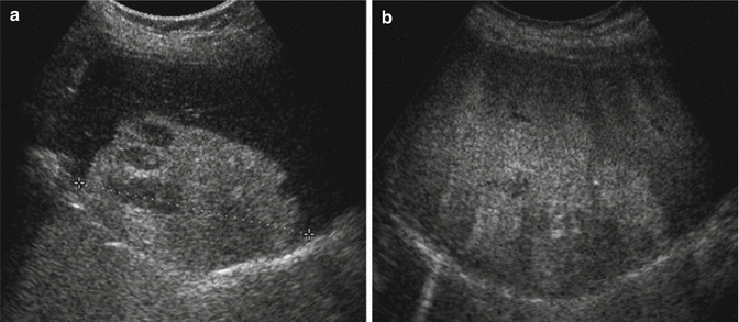
Fig. 5.1
Hepatic angiomyolipoma. (a) Right subcostal ascending US scan shows in segment VII a wide markedly hyperechoic lesion with regular margins (calipers) and some small hyperechoic area in the context. (b) After contrast medium injection, the lesion appears inhomogeneously hypervascular
5.2 Solitary Necrotic Nodule
The solitary necrotic nodule (SNN) is a rare benign tumor of uncertain origin often incidentally discovered. According to some authors, it represents a natural evolution of traumatic injury, infection by parasites, or a sclerosing hemangioma and can present as single or multiple lesions and is often misinterpreted as metastasis [7–9]. At microscopic examination, it can show different aspects: (1) an area of central coagulation necrosis often partially calcified, surrounded by a fibrous capsule in which inflammatory cells can also be evident; (2) coagulation necrosis mixed with liquefactive necrosis; and (3) multinodular fusion [10].
At US, SNN appears mainly hypoechoic and can show a “target” aspect with a central hyperechoic area and calcifications. According to literature, US aspect depends on the homogeneity of necrosis and the degree of dehydration of the lesion. Usually, it has a bilobed or polilobulated form, unusual for metastases, and grows in proximity to vascular structures. At color-Doppler, it shows no signal [9, 11]. After administration of USCA, SNN does not show substantial changes throughout the vascular phases, and as reported in literature, it shows mild or moderate enhancement at CT and MR [10, 12, 13].
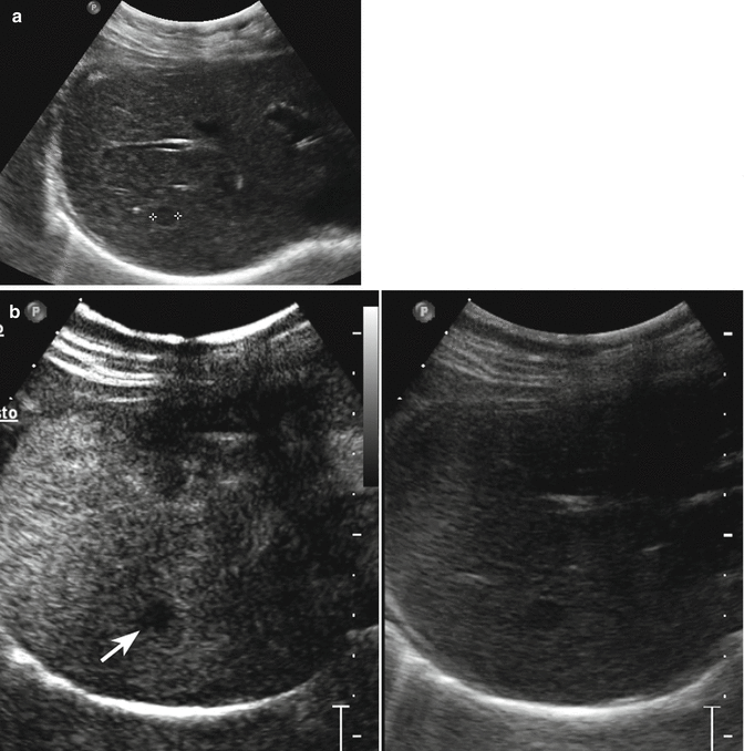
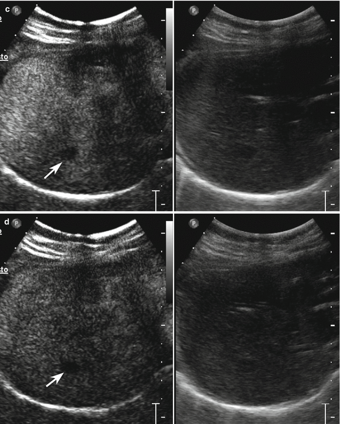
Fig. 5.2
Solitary necrotic nodule in a 66-year-old woman. (a) Oblique ascending right subcostal baseline image reveals a small homogeneous hypoechoic lesion sized 1 cm in the VII hepatic segment (calipers). (b–d) At CEUS, the lesion presents hypoechoic aspect throughout the vascular study (arrows)
5.3 Inflammatory Pseudotumor
The inflammatory pseudotumor (IP) is a benign rare lesion characterized by chronic infiltration of inflammatory cells and areas of fibrosis. It occurs more frequently in the lung whereas the incidence in the liver is very low (around 0.7 %) [14].
The pathogenesis is not clear. Infectious condition, autoimmune phenomenon, or systemic inflammatory response syndrome have been considered as possible pathogenesis [15]. IP can be associated with abdominal pain, vomiting, fever, weight loss, or increased inflammatory markers often directing toward a cancer diagnosis or be completely asymptomatic [16–18]. The final diagnosis, difficult also by means of CT and MR for the absence of peculiar imaging findings, is often obtained by biopsy or surgical resection [19].
At US, the lesion presents mainly hypoechoic, with well-defined margins and homogeneous or inhomogeneous echotexture due to the presence, in some case, of a central hyperechoic or anechoic portion. Usually at color- and power-Doppler evaluation, vascular signal is absent [20]. At CEUS, different patterns can be observed: (a) the lesion can appear constantly hypoechoic; (b) during the arterial phase, IP can show diffuse homogeneous hyperenhancement, diffuse heterogeneous hyperenhancement, peripheral rim-like enhancement, and isoenhancement appearing mainly hypoechoic during the extended portal-venous phase [21, 22].
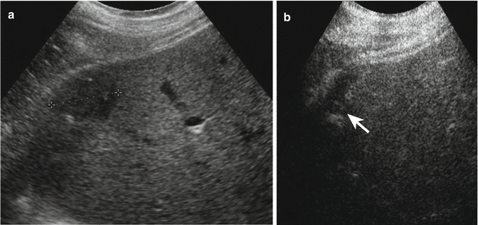
Fig. 5.3
Inflammatory pseudotumor in a 65-year-old man. (a) Oblique ascending right subcostal baseline image reveals a hypoechoic lesion sized 3 cm in the VI hepatic segment (calipers). (b) At CEUS, peripheral enhancing vessels are appreciable in the portal-venous phase, but the lesion remains hypoechoic (arrow)
5.4 Hemangiopericytoma (Lypomatous Subtype)
Hemangiopericytoma is a rare hypervascular tumor which can develop throughout the body in soft tissue and bone, arising from primitive mesenchimal cells located around the vessels called cells of Zimmerman. Hemangiopericytoma may occur at any age but is most common in fifth and sixth decades with the same frequency in men and women. Usually, this mass arises from the upper and lower extremities but, although infrequently, can develop in the liver too [23, 24].
Most commonly, it shows an intermediate grade of aggressiveness [23]. Anyway, large size (>5 cm), necrosis, increased cellularity, increased mitotic activity (>4 mitotic figures/10 high-powered fields), and cytologic atypia are considered prognostic negative features. So surgical removal is the first treatment with a 5-year survival rate of 70 %. Few data are reported in literature about US, CT, and MR imaging findings. It was described as a large, solitary, well-defined cystic lesion with a hypervascular solid component in the context. Speckled calcifications, necrotic and hemorrhagic areas, and cystic degeneration can occur, but these findings are not sufficient for the final diagnosis that is always surgical [25, 26].
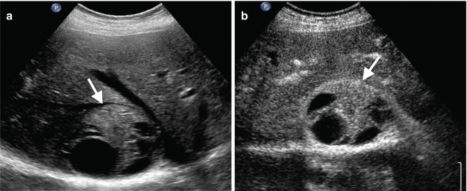
Fig. 5.4
Surgery-proven hemangiopericytoma (lypomatous variant) in a 68-year-old woman. (a) Right subcostal oblique ascending US scan shows a round, inhomogeneous 7 cm-sized lesion with well-defined borders in segment VII adjacent to the right hepatic vein (arrow). (b) After contrast medium injection, the lesion presents inhomogeneous aspect with some fluid areas in the context (arrow)
5.5 Extramedullary Intrahepatic Hematopoiesis
The extramedullary hematopoiesis (EH) is considered as a compensatory phenomenon to insufficient production of red blood cells by the bone marrow.
EH involves not only the liver but also other districts such as spleen, lymph nodes, chest, and kidneys. It is associated with severe anemia, congenital hemoglobinopathies, and acquired diseases such as leukemia, lymphoma, or myelofibrosis [27]. At US, the lesion shows well-defined margins, hypoechoic or hyperechoic appearance, and homogeneous or inhomogeneous echotexture due to the presence or not of fatty content [28]. Color Doppler can depict intralesional arterial vascularization [27, 29]. Usually, at CEUS, the lesion appears markedly hypervascular in the arterial phase with sustained but progressively decreasing enhancement in the extended portal venous phase as for focal nodular hyperplasia.
However, the rarity of the lesion and quite unspecific postcontrast findings make necessary biopsy or cytology for the definitive diagnosis in the majority of cases [30].
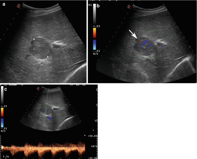
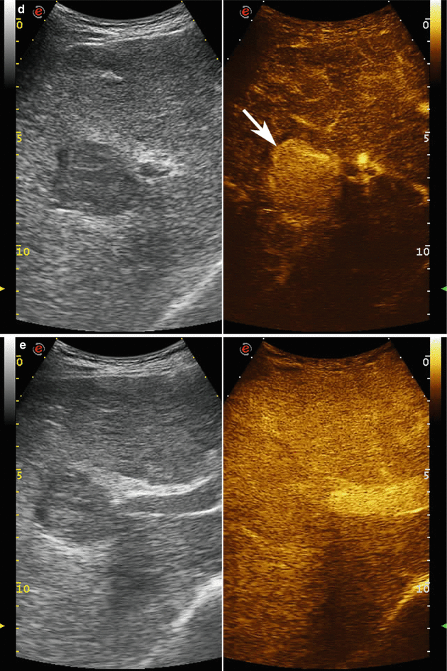
Fig. 5.5
Extramedullary erythropoietic hepatic nodule in a 53-year-old man affected by thalassemia. (a) Baseline US image shows a homogeneous hypoechoic lesion sized 4.5 cm in the VI–VII hepatic segment (calipers). (b, c) At color-pulsed Doppler evaluation, arterial vascular signal is evident within the mass (arrow). (d) At CEUS, the lesion appears highly and homogeneously hypervascular in the arterial phase (arrow) showing isovascular aspect with respect to the surrounding liver parenchyma during the extended portal-venous phase (e)
5.6 Hepatic Splenosis
Splenosis is considered as the heterotopic autotransplantation of splenic tissue, more frequently consequence of splenic trauma or surgery in the abdominal, pelvic, or thoracic cavity [31].
It commonly appears on the serosal surfaces of the intestine and mesentery, the omentum, the diaphragm, and the pelvis. In most cases, splenosis is asymptomatic; however, abdominal symptoms such as pain, recurring Felty syndrome, and intestinal obstruction have been reported [32, 33].
Related posts:


Stay updated, free articles. Join our Telegram channel

Full access? Get Clinical Tree


