| 7 | Normal Postoperative Appearances |
Restoring Intestinal Continuity
Following surgical resection of the colon—whether due to benign or malignant disease—the continence of elimination of feces must be restored. If continuity restores the natural pathway, i. e., so that evacuation occurs transanally, an anastomosis is necessary between resected segments. Continuity can be restored using end-to-end, end-to-side, or side-to-side anastomoses (Fig. 7.1).
 Ileocolic anastomosis. An ileocolic anastomosis is an anastomosis between the ileum and the colon (e.g., after a right hemicolectomy). This is generally an end-to-end (Fig. 7.2) or end-to-side anastomosis (Fig. 7.3). One can usually see a slight difference between the velvety, small intestinal mucosa and the smooth, shiny colonic mucosa. The transition is usually visible as a clear, smooth border and is only seldom slightly polypoid in appearance (Figs. 7.2, 7.3).
Colocolic anastomosis. A colocolic anastomosis is constructed between two colon segments (e.g., following resection of the sigmoid colon) and is usually an end-to-end anastomosis. A normal colocolic anastomosis appears as a smooth, whitish curvilinear scar that does not noticeably narrow the lumen (Fig. 7.4). Sometimes larger venous vessels can be seen near the anastomosis, but not intersecting it; occasionally suture remnants or metal staples (from the use of automatic sutures) can be seen on the anastomosis (Fig. 7.5).
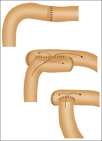
Abb. 7.1 Schematic illustration of options for restoring continuity following intestinal resection: end-to-end anastomosis (top), side-to-side anastomosis (middle), end-to-side anastomosis (bottom).
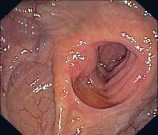
Abb. 7.2 Ileocolic end-to-end anastomosis.
Related posts:


Stay updated, free articles. Join our Telegram channel

Full access? Get Clinical Tree


