Normal Anatomy and Variants
Susan Hilton MD
Case 4-1
History: 41-year-old male with hematuria.
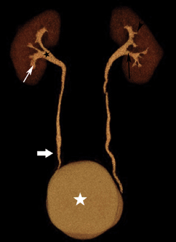 Figure 4.1 D (See color image.) |
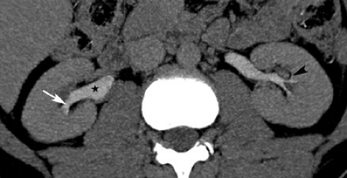
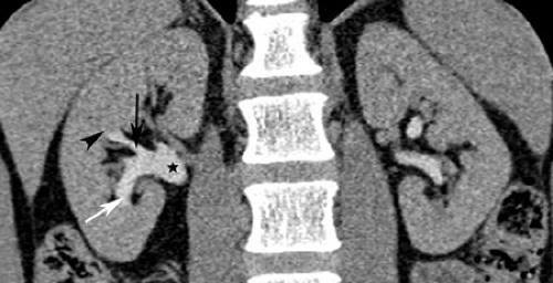
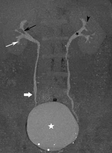
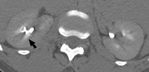
Findings: Axial image (A), coronal image (B), maximum intensity projection image (C), and volume-rendered image (D) from a normal CT urogram (See Color Fig. 4.1D.). The patient was also administered 10 mg of furosemide intravenously. The renal parenchyma, shown on the volume-rendered image in red, is normal in thickness. The calyces (white arrows) and fornices (black arrowheads) are normal. Other normal structures depicted include the infundibula (black arrows), renal pelves (black stars), ureters (thick white arrow), and bladder (white stars).
Diagnosis: Normal CT urogram
Discussion: The kidneys are retroperitoneal structures located within Gerota’s fascia. The superior one-third of the kidney is referred to as the upper pole, the middle one-third as the interpolar region, and the inferior one-third as the lower pole. The kidney is made up of lobes consisting of medullary pyramids that contain the collecting tubules and are surrounded by cortex except at the apex of the pyramids, at the papillae. Each papilla, the innermost portion of the medulla, projects into a calyx. The sharp-edged portion of each calyx extending around the papilla is called the calyceal fornix. The calyces join to form infundibula, which drain into the renal pelvis. Simple calyces are sharp and cup-shaped and drain one renal lobe; compound calyces, more common at the poles of the kidney, are complex in shape and drain several renal lobes (see Case 4-4). The renal pelvis is triangular, with its base in the renal sinus. The apex of the renal pelvis extends inferiorly to join the superior portion of the ureter and form the ureteropelvic junction. The ureters course inferiorly in the retroperitoneum, anterior to the psoas muscle, and cross the pelvic brim to enter the pelvis. The ureters then enter the bladder at an oblique angle to form the ureterovesical junction. (Case courtesy of Stuart G. Silverman, MD, Brigham and Women’s Hospital, Boston, MA.)
Case 4-2
History: 46-year-old female with hematuria.
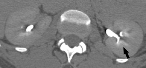
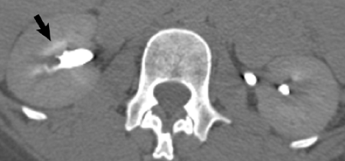
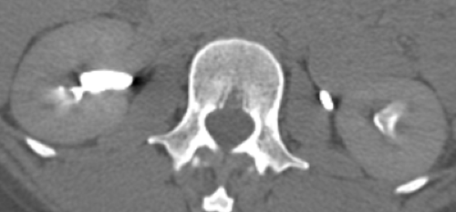
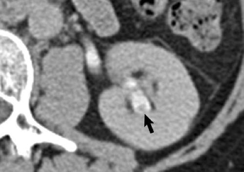
Findings: Excretory phase axial images (A, B, C and D) demonstrate dense radially arranged indistinct opacities in the renal papillae bilaterally (arrows).
Diagnosis: Normal papillary blush
Discussion: In healthy patients undergoing CT urography, visualization of indistinct opacities in the renal pyramids may be seen. This finding has been recognized on intravenous urograms for many years and is thought to be due to the normal concentration of contrast material in the medulla (see Case 11-12). The finding has been reported to be accentuated with low-osmolality contrast media because with the less hyperosmolar low-osmolality contrast media (compared to high-osmolality contrast agents) less fluid is excreted into the tubular lumen and, therefore, the concentration of contrast material is higher than with high-osmolality contrast material. Due to the superior contrast resolution of CT urography compared to intravenous urography, a normal papillary blush is displayed more frequently and more prominently. The “blushlike” appearance should be differentiated from the “brushlike” appearance of multiple, discrete, linear, papillary densities found in renal tubular ectasia (see Cases 4-10, 8-1, and 11-2).
Case 4-3
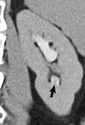
History: 55-year-old female with hematuria.
Findings: Axial (A) and coronal (B) images during the excretory phase of a CT urogram demonstrate an apparent small (4 mm) intraluminal filling defect (arrows) within a calyx of the lower pole of the left kidney.
Diagnosis: Prominent normal papilla
Discussion: CT urography may be used to differentiate a prominent normal papilla from a calyceal-filling defect due to a pathologic process. A prominent normal papilla has a central location within the surrounding calyx, a conical shape, and a smooth contour, as seen in this case (see Case 11-8). Also, in most instances, multiple papillae in the same kidney have the same appearance. Often, the diagnosis can be confirmed by viewing the collecting system in orthogonal planes. (Case courtesy of Stuart G. Silverman, MD, Brigham and Women’s Hospital, Boston, MA.)
Case 4-4
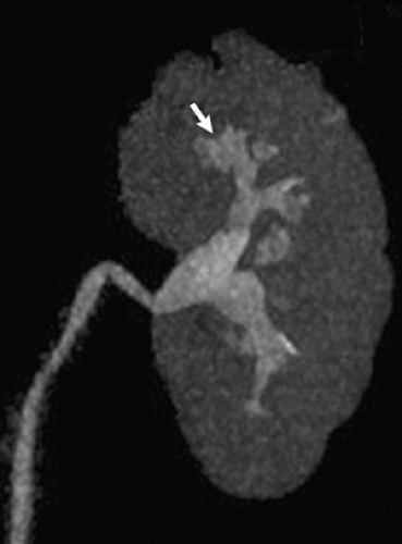
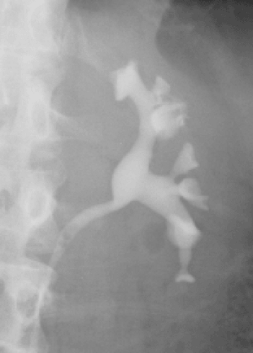
History: 56-year-old female with hematuria.
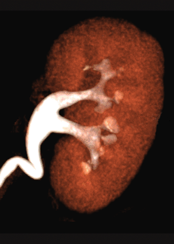
Findings: A compound calyx of the upper pole of the left kidney (arrow) is displayed on a volume-rendered coronal image of the left kidney (A) and on a left retrograde pyelogram (B).
Diagnosis: Compound calyx
Discussion: Embryologically, the kidney consists of multiple lobes. The collecting ducts of a renal lobe drain through the papilla of a medullary pyramid into a calyx. With simple calyces, each minor calyx empties into a single papilla, such that the number of calyces would equal the number of lobes if all calyces were simple. With compound calyces, multiple papillae empty into a single calyx. Compound calyces are most common in the upper pole and least common in the interpolar region of the kidney. Seventy percent of patients have one or more compound calyces. A compound calyx has also been referred to as a “T shaped” or “hammerhead” calyx. (Case courtesy of Stuart G. Silverman, MD, Brigham and Women’s Hospital, Boston, MA.)
Case 4-5
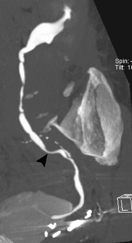
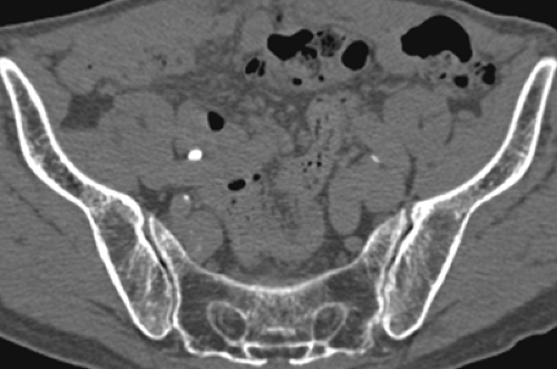
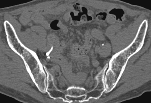
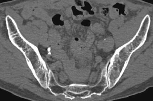
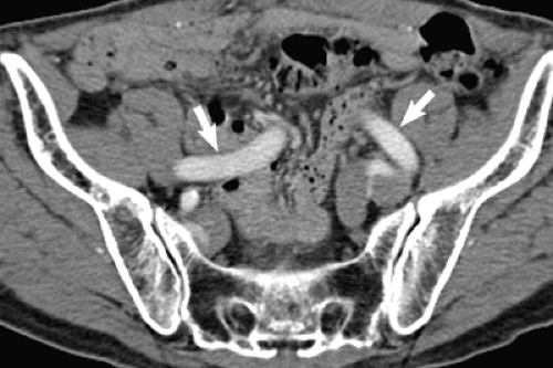
History: 77-year-old male with hematuria status post prostatectomy.
Findings: Curved planar reconstructed image (A), and axial images (B, C and D), during the excretory phase shows a narrowed right ureter (arrowhead). Axial image (E) during the arterial phase shows tortuous common iliac arteries (arrows). The ureter is narrowed at the same level as the calcified iliac artery (arrowhead). The right ureter just above the level of the iliac artery is of normal caliber (B), the ureter smoothly narrows as it crosses the tortuous artery (C), and the ureter resumes normal caliber just below the vessel (D).
Diagnosis: Narrowed right ureter due to adjacent atherosclerotic iliac artery
Discussion: The normal ureter is narrowed typically at three sites: the ureteropelvic junction, the point where the ureter crosses the common iliac vessels, and the ureterovesical junction. Narrowing and external impression of the ureter is more pronounced in the presence of atherosclerosis that causes ectasia and tortuosity of the iliac vessels. Vascular impressions may also be seen in the renal pelvis, where a normal, crossing renal artery branch may produce a bandlike or notchlike impression. Prior to the use of CT, this finding on IV urography was occasionally so problematic that angiography was needed to confirm the diagnosis. Today, vascular impressions on the urinary tract can be assessed fully with CT urography.
Case 4-6
History: 77-year-old male with hematuria.
 Get Clinical Tree app for offline access 
|
