Fig. 18.1
A transparent hood (D201-10704, Olympus, Tokyo, Japan) attached to the tip of the endoscope

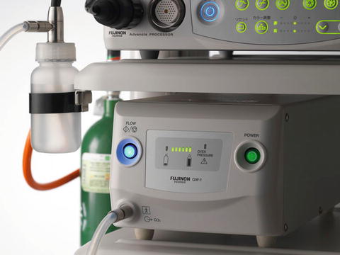
Fig. 18.2
CO2 insufflation device (GW-1, Fujifilm, Tokyo, Japan)
Desire for a Larger Accessory Channel
The therapeutic DBE with a 2.8 mm accessory channel (EN-450T5) can accommodate most of the accessory devices for endoscopic treatment, such as clip devices and balloon-dilation catheters. However, the insertion of these accessory devices can be difficult at times because the channel is tight and the endoscope shaft is long and often intricately looped. Moreover, suctioning of intestinal fluid or blood is almost impossible while accessory devices are in the channel. Therefore, a therapeutic DBE with a larger accessory channel has been long desired.
Development of New Therapeutic DBE (EN-580T)
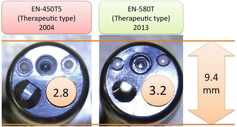
The distinctive feature of the double-balloon scope is the effective transmission of endoscopic control to the tip from the proximal endoscope shaft through the balloon overtube (Video 18.1). The transmission of the control does not rely on the stiffness of the shaft. Therefore, a thin and soft endoscope can be inserted and controlled in the deep small intestine. In order to intubate the soft and tortuous small intestine safely and effectively, the endoscope shaft should be thin and soft. Therefore, the manufacturer kept the same outer diameter of the endoscope even though the size of the accessory channel is increased to 3.2 mm. The new therapeutic EN-580T has a larger 3.2 mm accessory channel, but its outer diameter is the same at 9.4 mm (Table 18.1 and Fig. 18.3). As a result, this new model has improved intervention capability while maintaining the same ability to insert deeply into the small bowel.

Table 18.1
Specification of the double-balloon endoscopes
Diagnostic type | Therapeutic type | Short type | ||||
|---|---|---|---|---|---|---|
EN-450P5 | EN-580XP | EN-450T5 | EN-580T | EC-450BI5 | EI-530B | |
Outer diameter (mm) | 8.5 | 7.5 | 9.4 | 9.4 | 9.4 | 9.4 |
Accessory channel (mm) | 2.2 | 2.2 | 2.8 | 3.2 | 2.8 | 2.8 |
Working length (mm) | 2,000 | 2,000 | 2,000 | 2,000 | 1,520 | 1,520 |
Viewing angle (°) | 120 | 140 | 140 | 140 | 140 | 140 |
Minimum focus distance (mm) | 5 | 2 | 3 | 2 | 3 | 3 |

Fig. 18.3
Comparison of the accessory channel sizes between EN-450T5 and EN-580T
Features of EN-580T
Improved Interventional Capabilities
With the large accessory channel, insertion of accessory devices is easier because of less friction between the channel and the devices. This is especially notable for the balloon-dilation catheter because with the smaller channel there was significant friction at the tip of the catheter.
Endoscopic hemostasis for bleeding lesions is one of the more common therapeutic interventions performed during deep enteroscopy. Argon plasma coagulation, injection therapy, and clip placement are all available with DBE. When attempting control of hemostasis, it is important to identify the bleeding point and precisely apply the endoscopic treatment to the appropriate blood vessel. In order to identify the bleeding point, washing and suctioning the blood at the area of bleeding is very important. Once the bleeding point is identified, an accessory device for hemostasis is inserted through the accessory channel. However, because of the long shaft of the double-balloon scope, and the time it takes to insert the device, the bleeding point is often covered by blood before the device is fully inserted. In such cases, the bleeding point could be difficult to identify and withdrawal of the device is then required to wash the blood again. Using the new therapeutic EN-580T, water infusion for washing and suctioning is possible while keeping the hemostatic device in the channel because there remains enough space between the channel and the device. This feature makes the hemostatic procedure much easier and reliable.
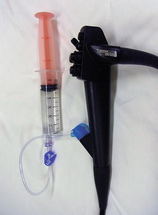
For the infusion of water through the accessory channel while keeping the accessory devices in place, BioShield irrigator (US Endoscopy, Mentor, OH, USA) is a useful tool (Fig. 18.4). Water can be infused through the irrigator using a syringe or a water pump. The BioShield irrigator is also useful for adding contrast medium during balloon dilation of an intestinal stricture. Balloon dilation is performed under fluoroscopy guidance after the stricture is visualized with contrast medium (Fig. 18.5a). However, after the first dilation with the balloon dilator, the contrast medium in the intestinal lumen often disappears distally down the intestine (Fig. 18.5b). In such cases, it was previously necessary with the 2.8 mm accessory channel to withdraw the balloon dilator to delineate the stricture again with additional contrast medium. Using the 3.2 mm accessory channel, however, contrast medium can be added through the BioShield while the balloon dilator is kept in the channel (Fig. 18.6a). Because the stricture can be delineated clearly again (Fig. 18.6b), the second dilation can be performed properly and safely (Fig. 18.6c).


