Modifying Sequence Parameters
This section describes the effects of modifying many of the user-defined parameters commonly encountered in abdominal and pelvic MRI. The primary (but not only) role of each parameter is listed, along with the effects of changing it in isolation. Although the effects of each parameter change described are typical, there are exceptions. For example, increasing the repetition time (TR) generally increases scan time. However, increasing the TR also allows data for more slices to be acquired during a multislice acquisition. This may eliminate the need for a second acquisition and, therefore, shorten overall scan time. Also, changing a scan parameter does not necessarily simultaneously result in all the effects described. For example, increasing slice thickness may result in increased anatomic coverage for the same scan time or the same anatomic coverage at decreased scan time.
Repetition Time
Primary effect: controls T1 contrast.
Increasing the TR:
Decreases T1 contrast.
Increases scan time.
Improves signal-to-noise ratio.
Increases the number of slices possible during a multislice acquisition.
Decreasing the TR has the opposite effect.
Note: For a gradient echo acquisition, T1 contrast is controlled by a combination of the TR and the flip angle.
Echo Time (see Figs. 1.7, 1.8)
Primary effect: controls T2 contrast.
Increasing the echo time (TE):
Increases T2 contrast (to a point).
Decreases signal-to-noise ratio.
Decreases the number of slices that can be obtained during a given TR.
Decreasing the TE has the opposite effect.
Note: Signal-to-noise decreases with increases in TE, because a longer TE allows more time for proton dephasing to occur. This results in a smaller net transverse magnetization vector and, therefore, less signal. For a gradient echo acquisition, the TE determines whether an in-phase or an opposed-phase image is produced.
Inversion Time
The effect of altering the inversion time (TI) for an inversion recovery sequence is not as straightforward as changing some of the other parameters discussed herein. For an inversion recovery sequence, a specific TI must be chosen to create the desired image contrast or to null signal from a specific tissue (e.g., fat). Increasing or decreasing the TI away from this value usually produces an undesirable result. For example, a short tau inversion recovery (STIR) sequence uses a specific TI (which depends on field strength) to null the signal from fat. Increasing this value may result in a water-suppressed image (because water has a longer T1 than fat and recovers through the null point at a later time). Decreasing the TI results in suppression of neither fat nor water.
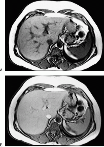
Flip Angle (Fig. 4.19)
Primary effect: controls image contrast.
Increasing the flip angle:
May result in more saturation of longitudinal magnetization (less signal).
Results in creation of more transverse magnetization (more signal).
Decreasing the flip angle has the opposite effect.
 FIG. 4.19. Effect on image contrast of changing flip angle from 80 to 30 degrees. A: T1-weighted spoiled gradient echo image of upper abdomen performed with flip angle of 80 degrees demonstrates excellent T1-weighted contrast with significant signal intensity difference between liver and spleen. B: Same sequence as (A) repeated with 30-degree flip angle demonstrates reduced image contrast.
Stay updated, free articles. Join our Telegram channel
Full access? Get Clinical Tree
 Get Clinical Tree app for offline access

|


