Fig. 6.1
MR images of a 63-year-old patient with prostate-specific antigen (PSA) progression from 3.8 ng/mL (2012) to 4.9 ng/mL (2013). (a) High-resolution axial T2-weighted fast spin-echo image showing a focal hypointense zone at the midperipheral gland in the postero-lateral location, without extracapsular extension: score 4. (b) Axial DWI image with b-value 3,000 mm2/s and (c) ADC map showing restricted diffusion phenomena of water molecules in the hypointense focus: score 5. (d) Colour DCE-MR map displaying avid enhancement of the hypointense zone with (e) type 3 signal intensity curve: score 4. (f) 1H-magnetic resonance spectroscopic imaging reveals in the zone of altered morpho-funcional MRI patterns, a choline-plus-creatine-to-citrate peak integral ratio greater than 1: score 5. The overall score of the lesion is PI-RADS 5: clinically significant cancer is highly likely to be present
Moreover, currently simultaneous imaging PET/MRI systems have been introduced in the clinical practice. PET/MR imaging provides combined structural, metabolic, and functional imaging information that can potentially affect patient management and outcome. Simultaneous acquisition of mp-MR and PET images with an appropriate radiotracer may be particularly valuable for identifying high-yield candidate biopsy sites that could reduce false-negative initial and repeated biopsies [23, 24].
6.4.1.1 T2-Weighted Imaging
T2W provides superior soft tissue contrast and clear delineation of prostatic zonal anatomy [25]. Most PC are low in T2 signal intensity against a background of high T2 signal intensity of the normal PZ, due to loss of normal glandular morphology with PC. However, low T2 signal intensity within the prostate is not always indicative of PC, as other benign conditions of the prostate, such as prostatitis, BPH, scars, or post-treatment changes (i.e. radiation and hormone ablation), post-biopsy haemorrhage, may have a similar low signal intensity. T2W can also assess whether the tumour is organ confined or extending beyond the prostate capsule. The detection of extracapsular extension (ECE) is quite important for preoperative staging because its presence upstages the patient to T3a stage, and thereby dictates a more aggressive treatment approach. On T2W, ECE usually appears as a direct extension of the tumour into the peri-prostatic fat, but ECE may not be obvious in all conditions; under such circumstances, secondary findings should be sought, including asymmetry of the neurovascular bundle, capsular obscuration or retraction and obliteration of the rectoprostatic angle. Seminal vesicle invasion (SVI) can be seen as a low signal defect within the normally high signal seminal vesicles (Fig. 6.2).
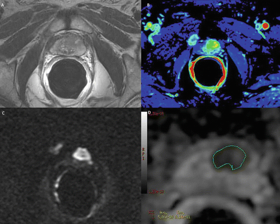
Fig. 6.2
MR images of a 75-year-old man with five negative TRUS-guided biopsies, PSA serum level of 32 ng/mL and PCa3 = 62. (a) High-resolution axial T2-weighted fast spin-echo image showing a focal oval-shaped hypointense lesion in the central zone with extension beyond of the anterior capsule. (b) Colour DCE-MR map displaying mild enhancement of the hypointense zone with. (c) Axial DWI image with b-value 3,000 mm2/s and (d) ADC map showing restricted diffusion phenomena of water molecules in the hypointense focus detected on T2-weighted images. Pathologic correlation after radical prostatectomy yielded central zone tumour
T2W alone is reported to have a wide range of Se and Spe for detecting PC (reported Se: 27–100 %; reported Spe 32–99 %), as well as for staging (reported Se 14–100 %, reported Spe 67–100 %) [26]. A recent study by Kim et al. [27] showed T2W alone at the high field strength (i.e. 3 T) revealing a detection accuracy of 80–90 % for tumour foci larger than 1.0 cm in diameter. However, for smaller tumours, T2W was far less accurate [28]. Concerning the evaluation of ECE, in a cohort of 108 patients, Bloch et al. [29] evaluated the role of T2W and DCE in staging PC and found an overall Se, Spe, PPV and NPV for ECE of 75, 92, 79 and 91 %, respectively. Calculi or clot within the seminal vesicles as well as unilateral atrophy can mimic SVI. However, combined use of T2W with DCE and DWI is helpful in this setting. In addition to lesion detection and local staging, T2W can also provide information about lesion size, which can be important in choosing the best treatment approach.
6.4.1.2 Diffusion-Weighted Imaging
DWI is based on an Echo-Planar sequence and depicts the diffusivity of water molecules along the three space directions within the tissue, which is decreased in the densely packed cellular structures of PC. DWI can be used to detect PC due to the lower ADC values of malignant tumours compared with non-cancerous prostate tissue. PCs within the PZ are hyperintense relative to a normal PZ on DWI and hypointense on ADC maps relative to a normal PZ. Recent evidence points to a correlation between a higher Gleason grade and lower diffusivity (low ADC) in PC [30]. Such a finding may, in the future, allow the non-invasive assessment of the histologic cancer grade. In general, at this time, an ADC below 1 × 10⁻³ mm2/s is regarded as suspicious for cancer [31]. The addition of DWI to standard imaging protocols dramatically improves the overall diagnostic efficacy of MRI for PC detection [32].
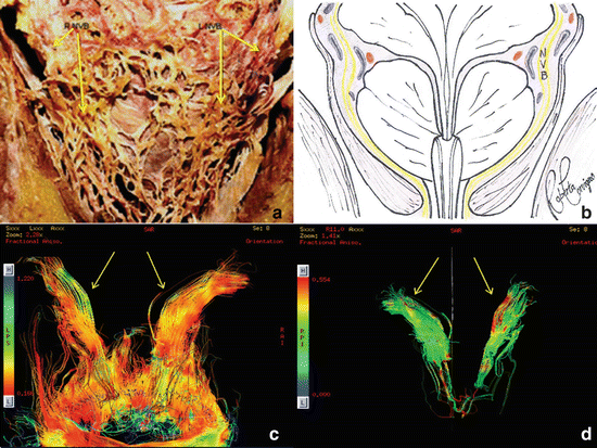
Diffusion tensor technique (DTI) is another Echo-Planar imaging technique that exploits the diffusivity of water molecules to map the orientation of sub-millimetric nerve fibres that unlike DWI highlights the diffusivity of water molecules along several space directions within the tissue. To date, DTI tractography has shown promising results in the visualisation of periprostatic nerve plexus. This information could be useful for guiding proper nerve-sparing surgery using an intra-fascial or extra-fascial robotic approach, thereby ensuring recovery of erectile function after RP [33].
6.4.1.3 Dynamic Contrast-Enhanced Imaging
DCE consists of the acquisition of sequential images using T1-weighted sequences during the passage of a contrast agent (such as low molecular weight gadolinium chelates) within the prostatic tissue. The pharmacokinetics of gadolinium-based contrast agents in the prostate produce different enhancement patterns in PC and benign tissues. The technique is based on the assessment of neoangiogenesis, which is an integral feature of tumours. DCE parameters can often be estimated both qualitatively and quantitatively. The main DCE parameters evaluated are: time to contrast peak (TTP), maximum slope of the contrast enhancement curve (MaxSlope), and area under the contrast enhancement curve (AUC). PC is characterised by a short TTP, high MaxSlope and a high AUC value [34]. However, abnormal enhancement patterns can be seen in BPH nodules and inflammation, making assessment of the central gland difficult [35]. Furthermore, smaller and low-grade tumour foci frequently do not show abnormal enhancement on DCE [36]. For the detection of tumours, DCE alone has a Se and Spe range of 46–96 % and 74–96 %, respectively. A recent work by Puech et al. analysed the performance of DCE in identifying and localising intraprostatic cancer foci in relation to cancer volume at histology. The Se and Spe of DCE for the identification of tumour foci of any volume were 32 and 95 %, respectively. For the identification of tumour foci >0.5 mL, the Se and Spe were 86 and 94 %, respectively, and the AUC was 0.874 [37]. The transitional zone (TZ) is a challenging area for tumour detection, as BPH nodules often show early and intense enhancement, much like tumours. However, similar to tumours in the PZ, TZ tumours also show early washout, which is unusual in BPH nodules. Nonetheless, it is important to interpret DCE of the TZ in the context of the T2W results. BPH nodules can often be distinguished by a clear capsular demarcation and a rounded appearance. It is uncommon for a BPH nodule to contain cancer.
DCE suffers from a relatively lower spatial resolution, so it must be combined with T2W to improve PC detection and local staging (AUC 95 % overall staging accuracy), compared with each technique alone [38]. DCE plays a key role in confirming a suspicion of SVI detected via T2W and/or DWI. The presence of early enhancement within a suspected seminal vesicle lesion strongly suggests invasion. Ogura et al. [39] reported that early, intense enhancement of the seminal vesicles has an accuracy rate of 97 % for SVI.
6.4.1.4 1H Spectroscopic Imaging
MRSI is a functional method used to assess prostate tissue metabolism. Each volume of interest (voxel) acquired in a 3D MRSI contains a metabolite spectra demonstrating the relative concentrations of metabolites such as citrate (Ci), creatine (Cr) and choline (Cho). PCs are characterised by increased levels of Cho and decreased levels of Ci, whereas normal prostate tissues contain high levels of Ci and relatively low levels of Cho and Cr. Thus, the increasing in the Cho + Cr/Ci and Cho/Ci are a marker for PC. Unfortunately, some benign conditions, such as prostatitis and postbiopsy changes, may also result in an increase of the ratio [40].
MRSI has been studied for several decades, and its joint use with T2W has been shown to aid in tumour volume estimation and tumour localisation. However, a recent prospective multicentre study carried out by the American College of Radiology Imaging Network to determine the benefit of combined ERC MRI and MRSI concluded that T2W alone and combined T2W-MRSI had similar accuracy in PZ cancer localisation (AUC, 0.60 vs. 0.58, respectively; P > 0.05) [41]. On the other hand, in a recent study, Selnaes et al. [42] found that combined T2W-MRSI performed better than T2W alone, with AUCs of 0.90 vs. 0.85. A principal advantage of the addition of MRSI to mp-MRI is its specificity [43].
6.4.1.5 Targeted-Biopsy Imaging Guided
Mp-MRI examination shows promising results in identifying suspicious foci of PC suitable for a rebiopsy in patients with persistently elevated PSA level and negative TRUS-guided biopsy.
Panebianco et al. in a prospective randomised trial conducted on 150 patients with initial negative TRUS-guided biopsy found that the combination of MRSI and DCE yielded 93.7 % Se, 90.7 % Spe, 88.2 % PPV, 95.1 % NPV and 90.9 % accuracy in detecting PC [44].
Bussetto et al. in a prospective study assessed the role of mp-MRI and urinary prostate cancer gene 3 (PCA3) test in identifying PC in 171 patients with negative prostate biopsy findings and a persistent high PSA serum level. The Se and Spe of the PCA3 test and mp-MRI was 68 % and 49 % and 74 % and 90 %, respectively, thus concluding that mp-MRI increases the accuracy and Se of the PCA3 test [45]. Sciarra et al. in a cohort population of 180 cases also show that in patients with a previous negative biopsy and persistently elevated PSA levels, the use of mp-MRI for indicating sites suitable for rebiopsy can significantly improve the sensitivity of the PCA3 test in the diagnosis of PC [46].
The data yielded by mp-MRI can be used to plan a transperineal MR-guided biopsy (MRGB). The in-bore approaches are exclusively MR based, using prebiopsy MRI to define the targets and real-time MRI to guide and control, with image confirmation, all steps of the procedure. The out-of-bore approaches use US to guide and control the procedure. The results reached by Penzkofer et al. [47] have shown that to date in-bore MRGB is very reliable and relatively easy, and the targeted approach has high yield with more positive lesions coming from ADC and DCE positive sites.
As in-bore biopsies require MR scanners, and thus valuable device time, they are associated with a higher organisational overhead as a result of the magnetic field hazards. Thus, in-bore MRI-guided prostate biopsy can be both time-consuming and expensive. However, it does offer the only method which can image the target and the biopsy needle within it prior to sampling, and thus the only true image-targeted biopsy. A proposed solution for this dilemma is the “out-of-bore” approach with fusion or registration of pre-procedural prostate MRI data to TRUS-guided biopsies, which combines the detection capabilities of MRI with the comparably easy set-up of TRUS [47]. The most straightforward approach for TRUS/MRI-guided biopsies is cognitive fusion, in which TRUS biopsies are performed knowing the localisation of MRI-suspicious lesions derived from peri-procedural MRI [47]. No specialised equipment is required other than an MRI scanner and a conventional TRUS biopsy device [48]. Despite the fact that cognitive fusion seems to improve biopsy protocols, more sophisticated devices have been developed for MRI/TRUS fusion that use different ways of registering the intraprocedural US coordinates to the MRI coordinates [47]. Pinto et al. [49] studied a group of 101 patients from three different risk categories derived from imaging aspects (low, moderate and high) on a TRUS/MRI system developed by Philips. All patients received 12-core standard systematic and MRI/TRUS fused prostate biopsies in the same setting. Cancer was detected in 27.9 %, 66.7 % and 89.5 % of the cases for the low, intermediate and high-risk groups, respectively. In this setting, MRI/TRUS fusion-guided prostate biopsies detected more cancer per core than the standard 12-core approach (20.6 % vs. 11.7 %). Sonn et al. [50] in a recent study concluded that TRUS/MRI biopsy is three times as likely to yield cancer diagnoses (21 % of performed targeted biopsies vs. 7 % of systematic biopsies), on a per patient basis, many cancers were detected by systematic biopsy alone (84 total positive diagnoses, 38 by both methods, 15 by MRI/TRUS alone and 31 by systematic TRUS-guided biopsy alone). Thus, the combination of systematic and TRUS/MRI-guided biopsy seems to be the key in the detection of more cancers.
6.5 How Can Advanced Imaging (mp-MRI) Change the Management of PC
Currently, the radiologists take substantial advantages from the new high technologies, especially mp-MRI. The benefits of mp-MRI include early detection of PC, the decreasing of unnecessary biopsies, the improving of biopsy accuracy, planning surgery, radiation treatment and focal therapies (such as high-intensity focused US, electroporation, focal brachytherapy and focal laser therapy). Thanks to DTI tractography technique, mp-MRI can identify the PC involvement of submillimetre fibres of periprostatic nerve bundles, thus playing a key role in the treatment planning (nerve sparing RP vs. non-nerve-sparing RP); moreover, displaying the anatomical course of periprostatic nerve fibres it is a reliable tool for guiding proper nerve-sparing surgery, thereby ensuring recovery of erectile function after RP [33] (Fig. 6.3).
 Early Diagnosis of Failure After Primary Treatment: Multiparametric MRI Versus PET-TC
 Prostate Cancer Units: How and Why
 Comparison Between a Multidisciplinary and a Monodisciplinary Approach to Prostate Cancer: Our 1-Year Experience
Intermittent Androgen Deprivation in the New Era: The Role of Urologist and Oncologist in a Multidisciplinary Team (MDT)
Comparison Between a Multidisciplinary and a Monodisciplinary Approach to Prostate Cancer: Our 1-Year Experience
Intermittent Androgen Deprivation in the New Era: The Role of Urologist and Oncologist in a Multidisciplinary Team (MDT)
 New Medical Strategies: The Role of Oncologist in an MDT
 Role of Pathology in the Multidisciplinary Management of Patients with Prostate Cancer


Related posts:
Early Diagnosis of Failure After Primary Treatment: Multiparametric MRI Versus PET-TC
Prostate Cancer Units: How and Why
Comparison Between a Multidisciplinary and a Monodisciplinary Approach to Prostate Cancer: Our 1-Year Experience
Intermittent Androgen Deprivation in the New Era: The Role of Urologist and Oncologist in a Multidisciplinary Team (MDT)
New Medical Strategies: The Role of Oncologist in an MDT
Role of Pathology in the Multidisciplinary Management of Patients with Prostate Cancer
Stay updated, free articles. Join our Telegram channel
Full access? Get Clinical Tree
