| 3 | Modern Endoscopic Techniques |
The use of video endoscopes has led to a significant increase in the quality of visualization compared with examinations performed using fiberoptic instruments. The charged-coupled device (CCD) chips used in modern video endoscopes now provide such high resolution that it is possible to detect even tiny details on the mucosal surface, which has led to significant advancements in detecting small and flat lesions, especially flat polyps. Yet, increasing resolution using video chips is only one means of increasing diagnostic yield in endoscopic procedures. Other advancements made over recent years, which will be discussed here, are:
 digital structural enhancement,
 magnifying or zoom endoscopy,
 chromoendoscopy,
 fluorescence endoscopy.
Digital Structure Enhancement
The higher resolution of modern video chips is accompanied by improved processing of the video signal by the computer to which the endoscope is attached. When the signal is transmitted to the image on the monitor, image processing (digital structure enhancement) allows surface structure details to be emphasized (Fig. 3.1). Extremely high resolution of detail is possible, especially when used with magnifying endoscopy and chromoendoscopy as discussed below.
Magnifying or Zoom Endoscopy
Magnifying or zoom endoscopes enable image enlargement to a point nearly comparable to intravital microscopic examination of the mucosa.
Automatic and electronic magnification systems. Enlargement can be achieved using a built-in powered lens system, computer-supported electronic magnification to produce a digitally enlarged image, or a combination of the two. Electronic magnification is technically simpler from an instrument standpoint and does not require moveable parts, but the enlarged image often appears “grainy.” The degree of detail in electronic magnification depends on the resolution offered by the CCD chip used, which limits the degree of magnification attainable. Mechanical enlargement, which uses moveable lenses, has the advantage of optical zooming (as opposed to mere digital approximation), similar to that of a microscope, with current magnification ranging from 100-150-fold. It has the disadvantage, however, of having to be integrated into the endoscope in a moveable lens system, including motorization (either manually with a linkage system or using a built-in miniature servo motor).
Discriminating detail. Extreme enlargement allows a high level of discrimination of the selected image. Yet, to achieve a sharp picture, the endoscope tip must be fixed very close to the mucosal surface being examined (focal distance). To do so, transparent caps are placed on the endoscope tip and then set on the mucosa to keep it at the correct distance from the instrument. Large vessels in the area or respiratory movements may cause movement artifacts, rendering zoom endoscopy impossible in isolated cases.
Magnification can reveal surface structure detail of even the smallest vessels (Figs. 3.2, 3.3), especially when combined with surface staining techniques (chromoendoscopy, see below).
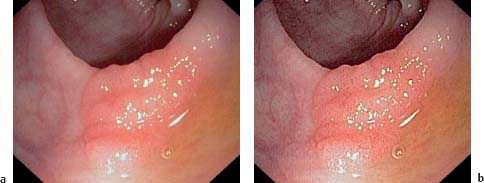
Fig. 3.1 Broadbased colonic adenoma.
a View without digital structure enhancement.
b View with digital structure enhancement. The surface structure can be seen more clearly.
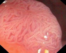
Fig. 3.2
Related posts:


Stay updated, free articles. Join our Telegram channel

Full access? Get Clinical Tree


