Minimally Invasive Splenectomy for Massive Splenomegaly

Chapter 21 Minimally Invasive Splenectomy for Massive Splenomegaly


The videos associated with this chapter are listed in the Video Contents and can be found on the accompanying DVDs and on Expertconsult.com.image


The first reported laparoscopic splenectomy was done by Delaitre and Maignien and published in 1992. Minimally invasive splenectomy, more commonly called laparoscopic splenectomy (LS), is now a standard surgical option for splenic disease; some authors believe that LS is the treatment of choice. Reported advantages of laparoscopic over open splenectomy (without splenomegaly) include shorter hospital stay, less narcotic administration, faster recovery, and a quicker return to employment. Although the most common indication for LS has been idiopathic thrombocytopenic purpura, many other benign or malignant hematologic diseases can benefit from this procedure. In a multicenter study on LS from the Italian Registry of Laparoscopic Surgery of the Spleen (IRLSS), malignancy was the underlying diagnosis in 32.8% of 676 patients.


Massive splenomegaly, defined as an organ larger than 20 cm in greatest dimension, is associated with various malignancies; is frequently seen in older, more physiologically frail patients; and can be associated with coagulopathy, anemia, thrombocytopenia, and perisplenitis. These preconditions, along with the risk for intraoperative hemorrhage and specimen extraction difficulties, made LS in the setting of massive splenomegaly a difficult challenge during the early experience, and massive organ size was a contraindication for LS. Most studies comparing LS in normal and enlarged (nonmassive) spleens have shown that LS for splenomegaly is associated with longer operative times, increased blood loss, more perioperative complications, longer length of stay, and higher conversion rates than LS for normal-sized spleens. Despite these findings, the general consensus has been that LS in experienced hands is preferable to open splenectomy for nonmassive splenomegaly.


Ongoing improvement in instrumentation and accrued experience have increased the rate at which LS is being done for massive splenomegaly, and the massively enlarged spleen is now a relative issue in the consideration of LS. Although LS for massive splenomegaly should not be used by all surgeons, in the appropriate hands this procedure is feasible for spleens of almost any size.


Operative indications


Benign splenic pathology seldom produces massive splenomegaly, with the exception of hemolytic anemia. Among malignancies, non-Hodgkin lymphoma (NHL) is by far the most common pathology associated with massive splenomegaly. Other associated malignancies include Hodgkin lymphoma (HL), idiopathic myelofibrosis, chronic lymphocytic leukemia, and hairy cell leukemia.


The patient with NHL may present with prominent retroperitoneal lymphadenopathy or hypersplenism without peripheral lymphadenopathy. Suspicious retroperitoneal lymphadenopathy is amenable to laparoscopic biopsy. The spleen is involved in 30% to 40% of patients with NHL. Treatment is directed at relieving the discomfort attributable to splenomegaly or preventing hematologic sequestration. Pancytopenia from the latter may prevent the administration of chemotherapy. Lymphoma confined to the spleen is rare (about 1% of NHL patients), but splenectomy may be curative. The patient with idiopathic myelofibrosis, chronic lymphocytic leukemia, or hairy cell leukemia may undergo splenectomy to treat symptomatic splenomegaly or hypersplenism, or both, similar to NHL.


In HL, the spleen typically is the first lymphoid basin that becomes involved below the diaphragm. The role of surgical staging has waned as imaging techniques and chemotherapy have improved, and staging is currently performed in about 5% of HL patients. A goal of noninvasive imaging in HL is to determine the presence of infradiaphragmatic disease. Staging laparoscopy, however, remains the most precise means of determining the presence and extent of abdominal involvement in patients with clinical Ann Arbor Stage I or II supradiaphragmatic HL; 20% to 35% of these patients have occult splenic or upper abdominal nodal involvement not detected by noninvasive imaging. An HL patient should be considered for staging laparoscopy (surface inspection, nodal sampling, liver biopsy) and splenectomy if the outcome of the procedure will influence subsequent therapy.


We consider most patients referred for elective splenectomy to be potential candidates for laparoscopic splenectomy. Contraindications to a minimally invasive approach include severe portal hypertension, uncorrectable coagulopathy, severe ascites, and most traumatic injuries. For a massive spleen that is less than 26 cm in greatest dimension, we proceed with LS in the absence of a contraindication. For a spleen that is larger than 26 cm in greatest dimension, we perform an open splenectomy through a left 14-cm subcostal incision. Similarly, other authors have suggested a cutoff dimension for laparoscopic splenectomy in the 27- to 30-cm range.


Hand-assisted laparoscopy or open splenectomy should be considered with extremely large spleens to avoid an intraoperative open conversion (per the guidelines of the European Association of Endoscopic Surgery), which is associated with an increased rate of postoperative morbidity. Other factors (e.g., morbid obesity, pancytopenia) may influence the surgeon to choose a more invasive approach. In the final analysis, the surgeon will have to judge whether to proceed with minimally invasive splenectomy for a massive spleen, a decision based on splenic size, relevant patient conditions, and the surgeon’s own experience and expertise with the technique.


Preoperative evaluation, testing, and preparation


The preoperative evaluation for a splenectomy patient should include sonographic examination to establish spleen size. Splenomegaly is defined as a maximal splenic diameter greater than 15 cm; a maximal diameter greater than 20 cm is indicative of massive splenomegaly (specimen weight, obtained after splenectomy, is not helpful in deciding surgical approach). Thin-slice spiral computed tomography (CT) should be used if additional anatomic information is needed or if malignancy is suspected. In autoimmune or hemolytic disease, thin-slice spiral CT scan may detect accessory spleens. For patients with malignant hematologic diseases, CT scanning can reveal lymphadenopathy in the splenic hilum, perisplenic inflammation, or splenic infarction, all of which can increase the difficulty of the procedure. CT scanning with three-dimensional reconstruction can add helpful information on possible anomalous vascular supply to the spleen so that the surgeon can appropriately modify the dissection. Magnetic resonance imaging also is useful, but is not routinely needed.


If the patient has thrombocytopenia, then treatment with prednisone (1 mg/kg/day, beginning 5 to 7 days before surgery) is recommended to achieve a preoperative platelet count higher than 50 × 109/L. If the platelet count is not increased with prednisone, then intraoperative platelet transfusion may be performed after division of the splenic pedicle. If the patient has autoimmune thrombocytopenia (not a common cause of massive splenomegaly), then administration of immunoglobulin is recommended. If the patient has anemia, then preoperative transfusion to raise the hemoglobin to 10 g/dL is advisable. Vaccination against meningococcal, pneumococcal, and Haemophilus influenzae type B pathogens is performed at least 15 days before elective splenectomy. Antibiotic prophylaxis (e.g., cefazolin or clindamycin) is administered 30 minutes before skin incision.


If the patient has a massively enlarged spleen, then preoperative splenic artery embolization may decrease both splenic size and the risk for intraoperative hemorrhage. We do not, however, use routine preoperative embolization because no clear advantage has been shown with this approach. Preoperative splenic artery embolization has been associated with severe pain, pancreatitis, and other ischemic complications. Furthermore, if lymphoma is suspected, then preoperative embolization may result in splenic necrosis, thus increasing the difficulty of pathologic diagnosis.


Patient positioning in the operating suite


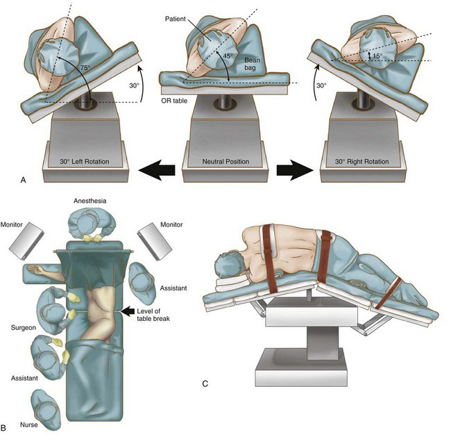
Laparoscopic splenectomy may be performed using a lateral decubitus, semi-decubitus, or supine approach, depending on surgeon preference, spleen size, patient characteristics, and the need for any concomitant procedures. The supine position provides access to the omental pouch, excellent visualization of the splenic hilum, and access to the rest of the abdomen if a concomitant procedure is planned. The decubitus position can facilitate bowel retraction away from the operative field, but full lateral decubitus alone is not recommended for a massive spleen because it will sag into the right upper quadrant, blocking the view of the hilum and making splenic manipulation extremely difficult. We prefer the semi-decubitus position for a massive spleen because the operating table can then be rotated to place the patient’s body in the supine or decubitus position (or anywhere in between), depending on surgeon need at any given point in the procedure (Fig. 21-1A).


Using a bean bag, foam wedges, or other stabilizing device, the patient is positioned with the left side up at about a 45-degree angle (Fig. 21-1B). Cushions are place under the knees and ankles of both legs. The right arm is extended onto an arm board, and the left arm is elevated and secured in a flexed position over the patient’s head. The patient is secured to the operating table with straps across the chest, hips, and legs to permit extreme rotation. The table is flexed to expand distance between the iliac crest and costal margin, and then reverse Trendelenburg tilt is applied (Fig. 21-1C). The monitors are placed on either side of the patient’s head. The surgeon and the camera operator stand on the patient’s right side, and the first assistant stands on the left, as shown in Figure 21-1B.


Positioning and placement of trocars


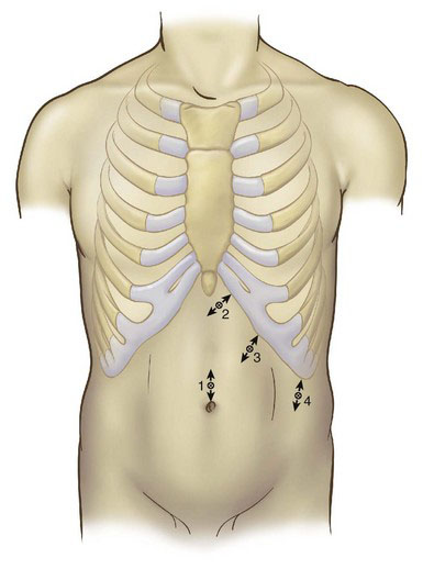
Four ports are used, as shown in Figure 21-2. A 10-mm camera port is inserted at the umbilicus using the open technique. If the patient is large or tall, or if the splenomegaly is moderate, then this initial port may be placed superior to or to the left of the umbilicus, or both. Under laparoscopic guidance, a 5-mm port is placed in the subxiphoid area, a 10-mm port is placed variably under the costal margin along the midclavicular line, and a 10-mm port is placed in the left axillary line, halfway between the costal margin and the iliac crest. The surgeon should adjust port placement to accommodate organ size. In massive splenomegaly, the subcostal ports are positioned 4 cm below the medial and inferior tip of the spleen, parallel to the left costal margin, yet still allowing the instruments to reach the diaphragm. Occasionally the most lateral port cannot be placed below the spleen; in such a case, the port should be placed as low as possible. Infiltration with local anesthesia before insertion of each trocar helps reduce postoperative pain and confirms the precise location of each port.


< div class='tao-gold-member'>

Stay updated, free articles. Join our Telegram channel

Jul 20, 2016 | Posted by in GASTOINESTINAL SURGERY | Comments Off on Minimally Invasive Splenectomy for Massive Splenomegaly

Full access? Get Clinical Tree

Get Clinical Tree app for offline access