Minimally Invasive Retroperitoneal Adrenalectomy

Chapter 34 Minimally Invasive Retroperitoneal Adrenalectomy


imageThe videos associated with this chapter are listed in the Video Contents and can be found on the accompanying DVDs and on Expertconsult.com.


The frequency of adrenalectomy has increased since 1980. This increase has been due in part to the increased quality and use of abdominal imaging. Incidentalomas account for a substantial number of adrenal lesions identified with abdominal imaging. Although most incidentalomas are nonfunctional and asymptomatic, some patients diagnosed with an incidentaloma ultimately will undergo an adrenalectomy because of changes in lesion characteristics or an increase in its size. Such changes in an incidentaloma raise the possibility of an adrenal neoplasm. The prevalence and incidence of adrenal masses requiring operative removal, however, is not well documented.


The increased frequency of adrenalectomy has been paralleled by an increase in the rate and types of minimally invasive adrenalectomy, including lateral transperitoneal, posterior retroperitoneal, lateral retroperitoneal, robotic, and needlescopic transperitoneal adrenalectomy. In general, the minimally invasive approach has been preferred for the removal of the small to moderate-sized adrenal mass since about 1995. The lateral transperitoneal approach has been the most common minimally invasive technique used in the United States. A less commonly used technique has been the minimally invasive retroperitoneal approach. Posterior retroperitoneoscopic adrenalectomy was popularized in Europe by Walz and coworkers and is the focus of this chapter. The advantages of this approach include direct access to the adrenal gland and the avoidance of intraperitoneal organs and adhesions.


Operative indications


The indications for posterior retroperitoneoscopic adrenalectomy are similar to other minimally invasive approaches, including nonfunctional incidentalomas, functional lesions producing Conn or Cushing syndrome, and pheochromocytoma. Adrenocortical carcinoma usually is considered a contraindication for minimally invasive adrenalectomy because of concerns of local invasion and incomplete resection. The relatively low incidence of adrenal carcinoma limits definitive assessment of these concerns.


Although tumors greater than 6 cm are considered a relative contraindication to minimally invasive adrenalectomy, larger lesions have been removed by the lateral transabdominal approach. An upper size limit for adrenal lesions has not been established for retroperitoneoscopic adrenalectomy. In the era of open surgery, a tumor size greater than 5 cm usually was considered a contraindication for open retroperitoneal adrenalectomy because of the confined operating space. For this reason, it seems reasonable to limit the use of retroperitoneoscopic adrenalectomy to lesions that are smaller than 5 cm.


Morbid obesity (body mass index [BMI] ≥40) complicates proper patient positioning and port placement for retroperitoneoscopic adrenalectomy; in particular, a thick layer of subcutaneous fat hinders port access to the posterior retroperitoneal space. In addition, the morbidly obese patient also is likely to have substantial retroperitoneal fat, which increases the difficulty of dissecting and identifying important structures. For these reasons, a BMI of 40 or greater is a relative contraindication for retroperitoneoscopic adrenalectomy, particularly early on in the surgeon’s experience with this technique.


Although the indications for adrenalectomy are the same for essentially all minimally invasive approaches, the retroperitoneoscopic approach offers some advantages compared with other methods to access the adrenal glands. In patients with prior abdominal surgery, retroperitoneoscopic adrenalectomy technique avoids intraperitoneal scar tissue and adhesions, reducing the risk for injury to the intra-abdominal organs. In addition, if the patient requires bilateral adrenalectomy, then repositioning is not necessary, as it would be if the patient were in the decubitus position.


Preoperative evaluation, testing, and preparation


Endocrine evaluation of functional disorders, such as Conn or Cushing syndrome, is performed in the standard fashion and is not reviewed here. The patient with pheochromocytoma should be treated with α-blockade for at least 2 weeks before surgery and should be hydrated during this period to prepare the cardiovascular system for adrenalectomy. Some pheochromocytoma patients also may require β-blockade for tachycardia. This latter treatment should never be started before α-blockade, however, because of the risk for unopposed α-adrenergic stimulation in a patient who already may have severe hypertension. Potassium deficits should be corrected and blood pressure controlled in the patient with an aldosteronoma. Inhibitors of steroidogenesis (e.g., ketoconazole, metyrapone) may be considered for patients with Cushing syndrome.


Although the physiologic changes associated with retroperitoneoscopy are not as well documented as those with pneumoperitoneum, some effects of increased pressure in the retroperitoneal space can be extrapolated from conventional laparoscopy, including decreased venous return from compression of the inferior vena cava, hypercarbia from carbon dioxide insufflation, and decreased renal perfusion. The patient with cardiac dysfunction, chronic obstructive pulmonary disease, or underlying renal disease who undergoes a retroperitoneoscopic procedure should receive the same preoperative optimization, intraoperative management, and postoperative monitoring of their comorbidities as would be done for a laparoscopic procedure.


Patient positioning in the operating suite


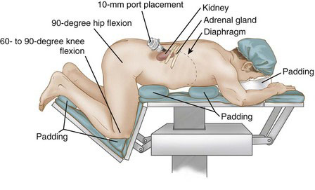
Retroperitoneoscopic adrenalectomy is performed with the patient in the prone jackknife position, as shown in Figure 34-1. The upper extremities are flexed at the elbows and pronated. The hips and knees are flexed at a 60- to 90-degree angle. It is important to flex the hips to expose the area between the posterior costal margin and the iliac crest and to permit adequate space for port placement. All pressure points, including the chest, hips, and knees, should be padded. The torso of the patient is kept parallel to the floor.


Positioning and placement of trocars


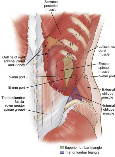
Posterior retroperitoneoscopic adrenalectomy can be performed using either a three-port or a single-port technique. With the three-port approach, the first (10-mm) trocar is placed 1 fingerbreadth below the tip of the 12th rib (Fig. 34-2). If fascia is encountered instead of muscle fibers, then it likely is the thoracodorsal (lumbodorsal) fascia, which invests the erector spinae muscles, meaning that the incision was too medial. If the thoracodorsal fascia is identified and the trocar is passed just lateral to this landmark, then the superior lumbar triangle likely will be traversed (see Fig. 34-2). The musculofascial layers in the region of the triangle are, from superficial to deep, the latissimus dorsi muscle, external oblique muscle, and transversalis fascia (Fig. 34-3). The superior lumbar triangle is the thinnest point of the posterior abdominal wall and is a potential location for incisional hernia. Ideally, therefore, the initial trocar should traverse lateral to this triangle. It often is difficult to ascertain the borders of this triangle, so this 10-mm port site should be closed securely at the procedure’s conclusion.
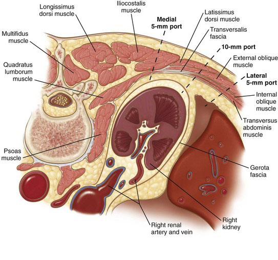


A properly placed 10-mm port-site incision should sequentially go through skin, subcutaneous fat, external oblique muscle, internal oblique muscle, transversus abdominis muscle, and transversalis fascia before reaching the retroperitoneal space (see Fig. 34-3). If the port is placed slightly cephalad and medial, then the surgeon will sequentially encounter skin, subcutaneous fat, tail of latissimus dorsi overlying the external oblique, external oblique, internal oblique, transversus abdominis, and transversalis fascia. Penetration through the transversalis fascia yields entry into the retroperitoneum, in particular the posterior pararenal space (see Fig. 34-2). Finger dissection can be performed through the port incision to increase the retroperitoneal working space. The 10-mm port then can be placed, followed by a 30-degree laparoscope; the latter can be used to bluntly dissect additional working space. Alternatively, a balloon dissector can be used to establish this space.


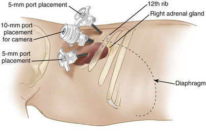
After a working space has been established, carbon dioxide is insufflated to a pressure of 20 to 25 mm Hg. Two additional 5-mm ports then are placed under laparoscopic visualization (see Fig. 34-2). The first is placed medial to the 10-mm port but lateral to the erector spinae muscles. Port traversal through the paraspinous muscles should be avoided because it is difficult to enter the working space and subsequently maneuver the port through this muscle mass. The second 5-mm port is placed lateral to the 10-mm port. The three ports should be aligned horizontally and spaced at least 3 cm apart to prevent interference (Fig. 34-4). The central (10-mm) port typically is used for the laparoscope. If a different dissection angle is desired, however, then a 5-mm laparoscope can be used through the medial or lateral port so that the working ports can be switched.


< div class='tao-gold-member'>

Stay updated, free articles. Join our Telegram channel

Jul 20, 2016 | Posted by in GASTOINESTINAL SURGERY | Comments Off on Minimally Invasive Retroperitoneal Adrenalectomy

Full access? Get Clinical Tree

Get Clinical Tree app for offline access