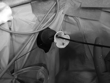
Fig. 23.1
Microperc set with the all-seeing needle (a) and flexible micro-optics (b) with the final vision of the percutaneous access after the needle removal (c)
23.2 Microperc and the Limitation of the Reduced Quality and Field of Vision
The necessity to employ a thin optics containing few fibres implies the generation of less light, thus a reduced illumination with a reduced image resolution of the working field. Additionally, the rigid optics has a limited field of vision, with no possibility to follow a mobile stone or escaped fragments during lithotripsy and no possibility to check all the calyces for residual fragments at the end of the procedure.
23.3 Microperc and the Limitation of the “Break and Leave” Principle
Microperc is based on the principle of “break and leave”, similarly to some RIRS and all ESWL. The lack of stone extraction after lithotripsy might be considered as a limitation factor, may cause renal colics postoperatively and also produce lower clearance rates [9]. Some urologists also succeeded in retrieving stone fragments for analysis using a basket catheter through the 8F microsheath [10].
23.4 Microperc and the Limitation of the High Renal Pelvic Pressures
If lithotripsy is carried out using a 200 μm laser fibre with adequate energy and frequency settings until complete disintegration of the stone, the issue of high renal pelvic pressures (RPP) for a prolonged time interval becomes relevant. From this point of view, Microperc suffers of the limitations of RIRS, working with a mean RPP of 33 mmHg and developing peaks as high as 170–328 mmHg [11].
Normal RPP is 5–15 mmHg, a 30–40 mmHg RPP for more than 10 min causes intrarenal reflux (pyelovenous, pyelolymphatic/pyelotubular, pyelointerstitial) with potential harmful consequences and forniceal damage takes place with 80–100 mmHg RPP [12]. Cadaveric studies revealed that with a 12/14F ureteral access sheath, even with high irrigation pressures, RPP remains within acceptable values [13]. On patients these data were also confirmed [14].
RPP generally remains low during PNL, if no mistakes like Amplatz malpositioning or anatomical problems like narrow infundibula occur [15]. During Microperc the irrigation system creates 50 mmHg RPP with a flow rate of 16 ml/min, and 100 mmHg RPP with 23 ml/min using saline when the optics is within the needle [6]. The question is whether this situation poses Microperc at increased risk of complications, especially in children [16].
23.5 Solving All Microperc Limitations: Micro-ECIRS
To our knowledge here, we report the first technical description of micro-ECIRS, a feasible and safe modification of the well-known one-step Microperc technique [17]. The idea of combining retrograde flexible ureteroscopy (with or without ureteral access sheath) with Microperc, with the patient in the Galdakao-modified supine Valdivia position (Fig. 23.2), is a proposal intended to solve most limitations of Microperc, also providing further advantages:
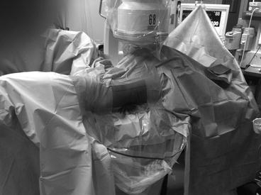
Fig. 23.2
Patient in the Galdakao-modified supine Valdivia position ready for micro-ECIRS
The morphology of the collecting system can be preliminarily examined, integrating the preoperative data from CT scans.
Stone features (mainly position, size, hardness) can be ascertained at the beginning of the procedure for an optimal strategic planning.
The best calyx to puncture can be chosen in real time, also examining its dynamic features with irrigation.
Access to the renal collecting system is achieved under fluoroscopic and Endovision control (Fig. 23.3) after preliminary ultrasound for controlling adjacent viscera and for identifying the correct inclination of the needle for the single access, avoiding as much as possible bleeding and multiple puncturing attempts.
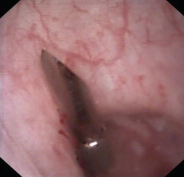
Fig. 23.3
Endovision application of the all-seeing needle
Migration of the stone into the ureter during initial irrigation or subsequent fragmentation is hindered.
Vision during lithotripsy is improved because of the retrograde illumination of the operating field (Fig. 23.4).
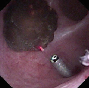
Fig. 23.4
The all-seeing needle with micro-optics and laser fibre inserted (retrograde flexible vision)
Vision during lithotripsy is also improved by the retrograde irrigation via the flexible ureteroscope.
Whether better vision reduces operating time remains to be determined [10].
Intrarenal pressures are concomitantly maintained low in presence of the ureteral access sheath, thus reducing resorption and uroseptic risk.
Thanks to the ureteral access sheath, sand from lithotripsy can be washed out retrogradely (Fig. 23.5).