Mesenchymal Tumors and Tumor-like Lesions


Mesenchymal Tumors and Tumor-like Lesions


Rare cases of benign mesenchymal neoplasms of the urinary tract have been described, although it is interesting to note that they are less common than their malignant counterparts. Primary sarcomas of the urinary bladder are rare, but are more common in males than females. The majority are of muscle origin and comprises less than 0.04% of all malignant tumors of the urinary bladder (1, 2, 3, 4). Myosarcomas may occur in any age group, but rhabdomyosarcomas (RMS) predominate in children while leiomyosarcomas (LMS) predominate in adults.


LEIOMYOMA

Leiomyoma is the most common of this group, exhibiting the same morphologic features seen at other sites (5, 6, 7) (Figs. 11.1, 11.2) (efig 11.1). On transurethral resection (TUR), there is a tendency to overlook the muscle as fragments of muscularis propria as the presence of circumscribed nodules cannot typically be appreciated. However, in contrast to the well-formed bundles of muscularis propria, leiomyomas are composed of large sheets of intersecting smooth muscle fascicles (Fig. 11.3). There is virtually no information on leiomyomas of the bladder with degenerative atypia (symplastic leiomyoma) (see “Leiomyosarcoma”).


LEIOMYOSARCOMA

LMS are more commonly seen in adults although there have been cases reported in children as well (1, 3, 5, 8). Rare cases have been reported subsequent to chemotherapy with cyclophosphamide (9, 10, 11, 12). The tumors are usually well circumscribed, may protrude into the lumen, and ulcerate the overlying urothelium. Size is variable although some may measure up to 20 cm. They occur more frequently in males than in females in a ratio of 2:1. Morphologic features are identical to those seen in other sites, including the presence of interlacing fascicles of spindle cells with variable amounts of eosinophilic cytoplasm with mild to marked nuclear atypia (Fig. 11.4) (efigs 11.2-11.7). Some tumors may have prominent myxoid change and others may have a prominent epithelioid morphology. The majority of
cases will demonstrate tumor necrosis and mitotic activity of 10 per 10 hpf or more. Although most smooth muscle tumors of the bladder are overtly either benign or malignant, there are some tumors with low cellularity yet with atypia. In the uterus, these lesions would be classified as benign, even if accompanied by readily identifiable mitotic figures. As there is no follow-up data in the literature for comparable lesions in the bladder, it may be more prudent to designate these lesions as “atypical smooth muscle tumors” describing their findings and stating that their biological behavior is unknown. For those lesions without mitotic figures, it can be added that it is likely that they will behave in an indolent fashion.


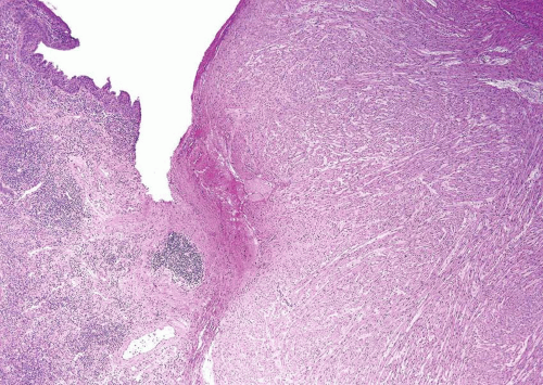
FIGURE 11.1 Leiomyoma.


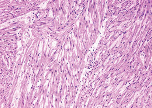
FIGURE 11.2 Leiomyoma (higher magnification of Figure 11.1).


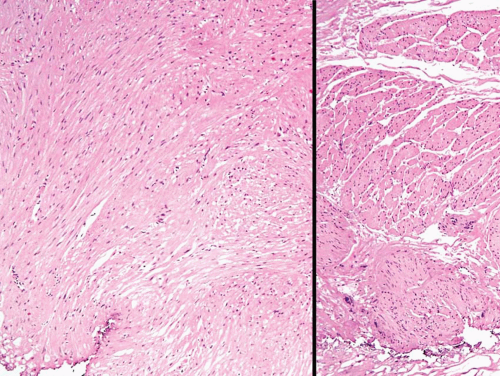
FIGURE 11.3 Leiomyoma composed of irregular sheets of smooth muscle (left) as opposed to well-formed discrete smooth muscle bundles of normal muscular propria (right).

Sarcomatoid carcinoma may mimic leiomyosarcoma, and must be excluded by adequate sampling of the specimen and appropriate histochemical, immunohistochemical, and electron microscopic studies. In fact, LMS of the bladder are so rare that this diagnosis should be made only after excluding all other possibilities, particularly carcinoma. LMS may express cytokeratin, but typically are composed of regular intersecting fascicles as opposed to the more haphazard growth pattern of sarcomatoid carcinoma. One must also rule out reactive spindle-cell lesions which may occur after local surgery or trauma. These reactive lesions are composed of spindle
cells that may express smooth muscle markers by immunohistochemistry but are of myofibroblastic origin (see below). Over two thirds of LMS will demonstrate immunoreactivity of both smooth muscle actin (1A4) and muscle-specific actin (HHF-35). Desmin will be positive in less than 50% of cases whereas epithelial membrane antigen is usually negative. Up to 50% of LMS may be immunoreactive for cytokeratins although focal (13). It has been suggested that caldesmon will be positive in LMS but negative in myofibroblastic lesions (14) (Table 11.1).


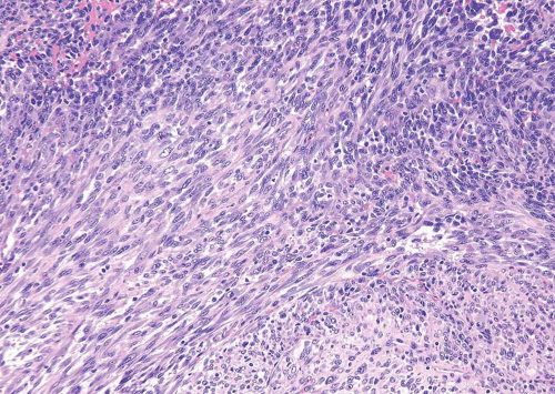
FIGURE 11.4 Leiomyosarcoma.


TABLE 11.1 Spindle Cell Neoplasms of the Bladder: Immunoreactivity


Antibody


RMS


LMS


SFT


IMT/PMP


SCa


Vim


+


+


+


+


+


SMA (1A4)


−/+


+


−/+


+/−


MCA (HHF-35)


+


+


−/+


+


Desmin


+


+/−


−/+


Myogenin


+


CD34


+


Bcl-2


−/+


+


−/+


−/+


EMA


+/−


CAM 5.2/


−/+


−/+


+/−


AE1/AE3 34BE12


+/−


ALK (IHC)


+/−


ALK (FISH)


+/−


RMS, rhabdomyosarcoma; LMS, leiomyosarcoma; SFT, solitary fibrous tumor; PMP, pseudosarcomatous myofibroblastic proliferations; IMT, inflammatory myofibroblastic tumor; SC, sarcomatoid carcinoma; Vim, vimentin; SMA, smooth muscle actin (clone 1A4); MCA, muscle common actin (clone HHF-35); EMA, epithelial membrane antigen (clone MC5); ALK, anaplastic lymphoma linase; +, positive in >75% of cases; +/−, positive in >50% of cases; −/+, positive in <50% of cases; −, negative in <5% of cases.


a Sarcomatoid carcinomas with mesenchymal differentiation may express muscle markers.


RHABDOMYOSARCOMA

In children, 20% to 27% of RMS arise in the genitourinary tract, the bladder, prostate, and paratesticular region being the most common primary sites in males and the bladder and vagina in females (15, 16, 17, 18) (Figs. 11.5, 11.6) (efigs 11.8, 11.9). The mean age at diagnosis for vesical RMS is 4 years. Most are embryonal RMS and exophytic (polypoid), with or without a “botryoid” component. Microscopically, the botryoid variant of RMS has a superficial
condensation of tumor cells, including strap cells and rhabdomyoblasts, located immediately beneath the urothelium (Figs. 11.7, 11.8). The underlying stroma is hypocellular and myxoid. In other polypoid tumors, the neoplastic cells are diffusely distributed throughout. A significant percentage of vesical RMS does not have an exophytic component and in these the tumor cells infiltrate the bladder wall diffusely (18). The spindle cell and alveolar variants of RMS may be rarely encountered. Typical rhabdomyoblasts and cross striations are seen frequently in exophytic RMS but rarely
seen in the spindle cell and alveolar types. Rare cases of vesical RMS have been described in adults and these may have embryonal, pleomorphic, or alveolar patterns (17). RMS should enter in the differential diagnosis of all spindle and myxoid lesions of the genitourinary tract in the pediatric age group. Tumor cells will be at least focally immunoreactive for desmin and myogenin, the latter in a nuclear distribution. Immunoreactivity for myoglobin is also diagnostic although it is positive in a minority of cases (Table 11.1).


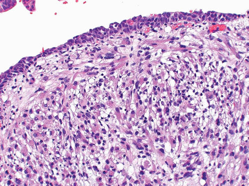
FIGURE 11.5 Rhabdomyosarcoma.


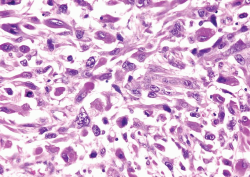
FIGURE 11.6 Rhabdomyosarcoma.


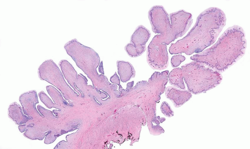
FIGURE 11.7 Botryoid variant of rhabdomyosarcoma.

These tumors may have remarkable overlap in morphology with small cell carcinoma, as the typical alveolar morphology may not be obvious and there may be greater degree of anaplasia (Fig. 11.9). Synaptophysin immunoreactivity further compounds this differential
although cytokeratin, myoD1, myogenin, and desmin should help confirm the diagnosis (19).


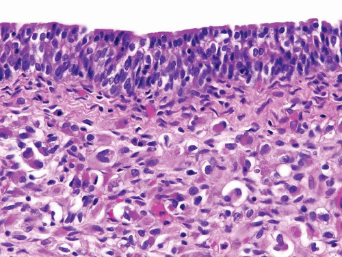
FIGURE 11.8 Cambium layer of botryoid variant of rhabdomyosarcoma (higher magnification of Figure 11.7).


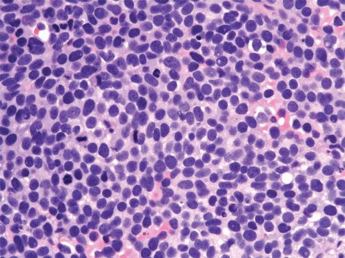
FIGURE 11.9 Rhabdomyosarcoma resembling small cell carcinoma.

In general, RMS have a poor prognosis in adults. Combination therapy with surgery and chemotherapy has greatly improved survival in the pediatric age group. Studies have suggested that exophytic RMS have a better prognosis than those that infiltrate the bladder wall diffusely (18). Patterns of embryonal RMS with a better prognosis include the spindle cell, botryoid, and tumors lacking diffuse anaplasia (criteria similar to Wilms tumors). Alveolar and pleomorphic variants are associated with a relatively poor outcome. Interestingly, treated tumors commonly exhibit morphologic evidence of “maturation” as evidenced by a greater number of myoblasts and cross striations (Fig. 11.10).


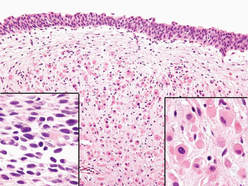
FIGURE 11.10 Rhabdomyosarcoma post-chemotherapy. Areas of the tumor showed mature rhabdomyoblasts (right inset) while others demonstrated cellular, more poorly differentiated sarcoma (left inset).


Jun 18, 2016 | Posted by in UROLOGY | Comments Off on Mesenchymal Tumors and Tumor-like Lesions

Full access? Get Clinical Tree

Get Clinical Tree app for offline access