| 10 | Malignant Tumors |
 Definition and Classification
Most colorectal carcinomas are adenocarcinomas, which developed from colorectal adenomas (adenoma-carcinoma sequence). Less than 2% of malignancies are nonepithelial malignancies (lymphomas) or colon metastases of another primary tumor. Adenocarcinomas account for the vast majority of carcinomas in the colon (85%) and rectum (90%); mucinous adenocarcinomas account for only ca. 13% of carcinomas in the colon and ca. 8% of carcinomas in the rectum. Table 10.1 gives an overview of the histological classification of primary colorectal tumors.
The degree of differentiation of adenocarcinomas is described by a grading system of the UICC, whereby the degree of differentiation or malignancy is usually divided into four grades:
 G1 = highly differentiated
 G2 = moderately differentiated
 G3 = poorly differentiated
 G4 = undifferentiated.
Signet-ring cell carcinomas are classified as G3; undifferentiated, small-cell carcinomas as G4. The TNM staging system (UICC 1993) is used especially for staging colorectal carcinomas in terms of extent of the local tumor, spread to lymph nodes, and potential distant spread (metastasis).
 Clinical Picture and Clinical Significance
Epidemiology. Incidence of colorectal carcinoma is generally high in western industrialized nations. In Germany, the rate among men is 41 per 100000 and among women it is 52 per 100000. For both men and women colorectal carcinomas are the third most common carcinoma. Frequency of disease rises steadily with increasing age, the maximum being around 75 years of age. Epidemiological studies have shown that in addition to genetic causes, there is also an association between environmental factors and the development of colorectal carcinomas. A diet rich in animal fat and low in fiber promotes the development of colorectal carcinomas. Further risk factors include chronic inflammatory bowel diseases (especially ulcerative colitis), colon adenomas, family history, prior ureterosigmoidostomy, and hereditary syndromes (familial adenomatous polyposes, hereditary nonpolyposis colorectal cancer).
Symptoms. About 60% of all colorectal carcinomas are located in the colon and around 40% in the rectum. Of those located in the colon, most (60%) are found the sigmoid colon, followed by the ascending colon. In 4-6% of patients, two or more synchronous carcinomas are found in the colon and rectum (1). The main symptom of colorectal carcinoma is the presence of blood and mucous in stool, though symptoms of right-sided colon carcinomas initially manifest themselves indirectly as iron-deficiency anemia resulting from chronic occult blood loss. Changes in bowel habits, unintended weight loss, tenesmus, and diarrhea are further typical symptoms that may indicate colorectal carcinoma. Around 10% of all colorectal carcinomas are diagnosed in emergencies related to an obstruction or perforation (1).
Epithelial tumors | Nonepithelial tumors |
|---|---|
|
|


Treatment strategy. Treatment strategies for colorectal carcinoma depend on the pathological tumor stage. The primary treatment for all colorectal carcinomas is surgical resection, which can also be indicated for already metastasizing disease, for example, in order to prevent bowel obstruction (ileus) and stenosis. Additional adjuvant and neoadjuvant therapies include chemotherapy and radiation according to tumor stage; for rectal carcinomas, further treatment is determined by pre-operative endosonographic T-staging (TNM staging). For metastasizing disease, chemotherapy may be a palliative treatment option.
 Diagnosis
Diagnosis of colorectal carcinoma
 family history,
 clinical examination (rectal palpation),
 laboratory parameters (CEA),
 total colonoscopy (with biopsy sampling),
 double-contrast enema examination of the colon, if necessary (if complete endoscopy is not possible),
 virtual colonoscopy (?),
 sonography of liver and abdomen,
 if necessary, abdominal computed tomography (CT)/ magnetic resonance imaging (MRI) of liver,
 thoracic radiography,
 endosonography, MRI with endorectal coil (rectal carcinoma).
 10.1 Macroscopic growth form of colorectal carcinoma: ulcerative tumor
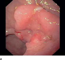
a Rectal carcinoma. Bowl-shaped ulceration, gray-yellow hue.
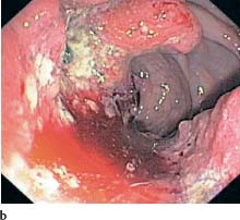
b Ulcerated colon carcinoma with inflammatory margin and petechiae.
Diagnosis is based on family history and clinical findings. Family history is especially important with regard to hereditary poly-posis syndromes and for patients with a history of colorectal adenomas and carcinomas in the family. Digital rectal examination is especially important for clinical findings, as 40% of all colorectal carcinomas are located in the rectum.
Role of endoscopy.


Stay updated, free articles. Join our Telegram channel

Full access? Get Clinical Tree


