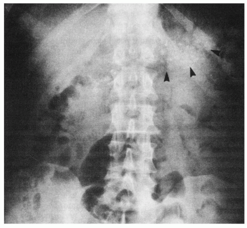
Malabsorption


Malabsorption


The term malabsorption connotes the failure to absorb or digest normally one or more dietary constituents. Patients with malabsorption often complain of diarrhea, and sometimes the distinction between malabsorption and diarrhea of other causes (see Chapter 29) initially is difficult. For example, patients with primary lactase deficiency fail to absorb a specific dietary constituent, lactose, and a watery, osmotic diarrhea develops. However, most patients with malabsorption present with a syndrome characterized by large, loose, foul-smelling stools and loss of weight. On additional study, it is found that they cannot absorb fat and often carbohydrate, protein, and other nutrients also. 
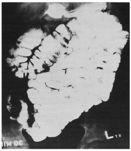
Table 31-1 indicates that a wide variety of disorders of the organs of digestion can cause malabsorption or maldigestion.


I. DIAGNOSTIC STUDIES.

Before discussing the disorders that may cause malabsorption, it is useful to review several of the diagnostic studies that are available to aid in evaluating patients with this condition. The number and order of diagnostic studies used depends on the clinical signs and symptoms of the patient.


A. Blood tests.

The hemoglobin and hematocrit levels may identify an anemia that accompanies malabsorption. A low mean cell volume (MCV) may be found in iron deficiency, whereas a high MCV may result from malabsorption of folate or vitamin B12. Serum levels of liver enzymes, protein, amylase, calcium, folate, and vitamin B12 may be abnormal and should be ordered.


TABLE 31-1 Disorders That May Cause Malabsorption or Maldigestion of One or More Dietary Constituents


Digestive disorder


Examples


Pancreatic exocrine insufficiency


Chronic pancreatitis


Pancreatic carcinoma


Bile acid insufficiency


Small-bowel bacterial overgrowth


Crohn’s disease of the terminal ileum


Small-bowel disease


Mucosal disorders


Celiac sprue


Collagenous sprue


Tropical sprue


Whipple’s disease


Radiation enteritis


Ischemic disease


Intestinal lymphoma


Regional enteritis


(Crohn’s disease)


Amyloidosis


Specific absorptive defects


Primary lactase deficiency


Abetalipoproteinemia


Lymphatic disorders


Intestinal lymphangiectasia


Mixed defects in absorption


Zollinger-Ellison syndrome


Postgastrectomy disorders


Figure 31-1. Plain x-ray film of the abdomen in a patient with extensive calcification of the pancreas (arrows) and pancreatic insufficiency.


B. Radiographic studies


1. Plain films or computed tomography scan of the abdomen

may show calcification within the pancreas, which indicates chronic pancreatic insufficiency (Fig. 31-1).


2. A barium examination of the upper gastrointestinal tract,

including the small bowel, usually is one of the first diagnostic studies in the evaluation of malabsorption syndrome. Often the findings are nonspecific. The bowel may be dilated and the barium diluted because of increased intraluminal fluid. A more specific finding is thickening of the intestinal folds caused by an infiltrative process, such as lymphoma, Whipple’s disease, or amyloidosis. The narrowed, irregular terminal ileum in Crohn’s disease is virtually diagnostic (Fig. 31-2), although lymphoma and other infiltrative disorders also must be considered. Diverticula, fistulas, and surgical alterations in bowel anatomy also may be evident.


C. Fecal fat determination.

Malabsorption of fat (steatorrhea) is common to most malabsorptive conditions (Table 31-2). Patients should ingest at least 80 g of fat per day to obtain reliable interpretation of qualitative or quantitative fat determination. Mineral oil and oil-containing cathartics should be avoided.


1. Qualitative screening test.

The Sudan stain for fecal fat is easy to perform and reasonably sensitive and specific when interpreted by an experienced person. A small amount of fresh stool is mixed thoroughly with normal saline or water on a glass slide. A drop of glacial acetic acid is added, and the slide is heated to hydrolyze the fatty acids from the triglycerides in the stool. The Sudan stain is then added. Increased stool fat is indicated by abnormally large or increased numbers (> 100/40 × field) of fat droplets.


2. The quantitative determination of stool fat

is more accurate than qualitative screening, but the collection of stool often is disagreeable to patients, family, and nursing personnel. The stool is collected over 72 hours in a large sealed container, which can be enclosed in a plastic bag and refrigerated to contain unpleasant odors. Most normal people excrete up to 6 g of fat per 24 hours on a diet that contains 80 to 100 g of fat. Stool fat in excess of 6 g per 24 hours can result from a disorder of fat digestion at any stage, including pancreatic insufficiency (decreased lipase), bile acid insufficiency, mucosal disease, or lymphatic obstruction.


Figure 31-2. Small-bowel x-ray series in a patient with Crohn’s disease. Note the narrowed, irregular contour of the terminal ileum and cecum (arrow). (From Eastwood GL. Core Textbook of Gastroenterology. Philadelphia: Lippincott Williams & Wilkins; 1984:102. Reprinted with permission.)


D. Pancreatic function tests


1. Collection of pancreatic secretions from the duodenum.

The volume of pancreatic secretion and the content of bicarbonate and enzymes can be measured by collecting pancreatic secretions from the duodenum after stimulation of the pancreas with secretin or with a test meal. Pancreatic insufficiency or carcinoma of the head of the pancreas, which partially obstructs the pancreatic duct, may be detected by this means. For example, bicarbonate concentrations less than 90 mmol/L suggest pancreatic insufficiency. However, pancreatic secretory tests are performed so infrequently in most gastrointestinal laboratories that the results may be unreliable.


2.

The bentiromide test is a test of pancreatic exocrine function that does not require duodenal intubation. The chemical name of bentiromide is N-benzoyl-L-tyrosyl-P-aminobenzoic acid. The test is performed by administering a single oral dose of 500 mg of bentiromide after an overnight fast and the urine is then collected for 6 hours. The pancreatic enzyme chymotrypsin cleaves the molecule within the lumen of the small intestine, releasing paraaminobenzoic acid (PABA). The PABA is absorbed and excreted in the urine. Less than 60% excretion of PABA suggests pancreatic insufficiency, although mucosal disorders, renal disease, severe liver disease, and diabetes also can cause low PABA excretion.


3. Radiographic studies.

Although computed tomography of the abdomen (see Chapter 9) and endoscopic retrograde cholangiopancreatography (ERCP) (see Chapter 5) do not measure pancreatic function directly, abnormalities such as
dilated or strictured ducts, calcification, and pancreatic masses can imply pancreatic disease.


TABLE 31-2 Expected Diagnostic Findings According to Cause of Malabsorption


Diagnostic Test


Disorder


Small-bowel x-ray


Fecal fat


Xylose tolerance test


Schilling test


Small-bowel biopsy


Pancreatic exocrine insufficiency


Abnormal


Severe steatorrhea


Normal


May be abnormal


Normal


Bile acid insufficiency


Normala


Mild-to-moderate steatorrhea


Normala


Normala


Normala


Small-bowel mucosal disease


Abnormal


Mild-to-severe steatorrhea


Abnormal


Usually normalb


Abnormal


Lymphatic disease


May be abnormal


Mild steatorrhea


Normal


Normal


Abnormal


a May be abnormal in bacterial overgrowth.

b Abnormal if terminal lieum is involved.


E. Bile acid breath test.

Conjugated bile acids that are secreted into the duodenum are resorbed with about 95% efficiency in the terminal ileum. If radiolabeled [14C]-glycocholate is given orally to a healthy person, about 5% of it enters the colon and undergoes bacterial deconjugation. The carbon dioxide (14CO2) derived from the glycine is absorbed and excreted by the lungs and can be measured in expired air. Bacterial overgrowth in the small intestine promotes earlier bacterial deconjugation of the [14C]-glycocholate, and consequently, a larger amount of 14CO2 is measured in the breath. Similarly, disease or resection of the terminal ileum allows more bile acids to pass into the colon and undergo bacterial deconjugation, resulting in an increase in expired carbon dioxide.

Jun 11, 2016 | Posted by in GASTROENTEROLOGY | Comments Off on Malabsorption

Full access? Get Clinical Tree

Get Clinical Tree app for offline access