CHAPTER 4 MAJOR HEPATIC RESECTION: LEFT LIVER
LEFT HEPATECTOMY*
INFLOW CONTROL
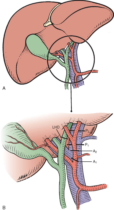
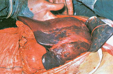
Dissection at the base of the umbilical fissure exposes the left portal vein. It should be controlled at this point and not at its origin from the main portal venous trunk. If the caudate lobe is to be removed together with the left lobe, the vein is secured below the takeoff of the caudate branches. If the caudate lobe is to be preserved, the vein is divided distal to the takeoff of the caudate branch (see Fig. 3-14). The left hepatic duct usually is easily identified just above and behind the left portal vein and can be encircled with a suture and divided as it curves into the umbilical fissure. These steps are illustrated in Fig. 4-1. A line of demarcation in the Glissonian plane extending from the gallbladder fossa to the left of the vena cava is now visible on the liver surface (Fig. 4-2) and is marked with a diathermy.
OUTFLOW CONTROL
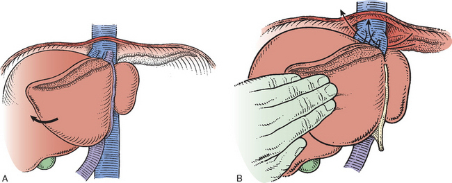
The left lobe of the liver (segments II and III) is now mobilized from the diaphragm by division of the left triangular ligament from its tip up to the lateral margin of the suprahepatic inferior vena cava (IVC) (see Fig. 2-18). The upper surfaces of the left and middle hepatic veins, or of their common trunk and of the suprahepatic IVC, are displayed (see Fig. 2-16) (Czerniak et al., 1993). In essence, the veins are cleared below the diaphragm, and their junction with the vena cava is displayed. The left lobe of the liver (segments II and III) is turned to the right (Fig. 4-3). The gastrohepatic ligament is fully divided, and the ligamentum venosum is identified. The ligamentum venosum is divided close to its entry into the left hepatic vein. This division opens up a triangular tunnel, the sides of which are the left hepatic vein anteriorly, the anterior surface of the vena cava posteriorly, and the upper surface of segment II inferiorly. This tunnel is developed carefully, and an instrument is passed beneath the left and middle hepatic veins to emerge anterior to the vena cava between the middle hepatic vein and the right hepatic vein (see Fig. 4-3).


Stay updated, free articles. Join our Telegram channel

Full access? Get Clinical Tree


