(1)
Department of Endoscopy, Fukuoka University Chikushi Hospital, Fukuoka, Chikushino, Japan
Summary
Basic microanatomical findings
Gastric body/fundic mucosa
V: Regular honeycomb-like SECN pattern with regular CV pattern present
S: Regular oval crypt opening pattern
Gastric antral mucosa
V: Regular coil-shaped SECN pattern but regular CV pattern absent
S: Regular groove-like crypt opening pattern
Keywords
CapillaryCrypt openingMagnifying endoscopyNormalStomachCollecting venule4.1 Explanation
The magnified endoscopic (ME) findings of the gastric mucosa, with no pathological changes such as Helicobacter pylori (H. pylori) infection, show a completely different pattern in the gastric body/fundus and antral regions [1, 2]. Interpretation of the normal gastric mucosa as seen using magnifying endoscopy first requires an understanding of how we visualize the microanatomical structures. In this chapter, I will explain the findings of magnifying endoscopy using WLI with indigo carmine dye spraying.
In the normal gastric mucosa, the basic anatomical constituents used to interpret the microvascular architecture (V) are the subepithelial capillary network (SECN) and subepithelial collecting venules (CV). Similarly, the basic anatomical constituents used to interpret the microsurface structure (S) are the crypt openings and gastric sulci.
4.2 Gastric Body and Fundic Mucosa
4.2.1 Microvascular Architecture (V)
In the normal gastric body and fundus (gastric fundic gland mucosa) (Fig. 4.1a), the microvascular architecture (V) shows a regular honeycomb-like SECN pattern. A polygonal closed loop of subepithelial capillaries surrounds each gastric pit, each loop anastomosing with its neighbor to form a honeycomb-like network beneath the epithelium. This capillary network converges on a CV somewhat greater in diameter than the capillaries (Fig. 4.1b).
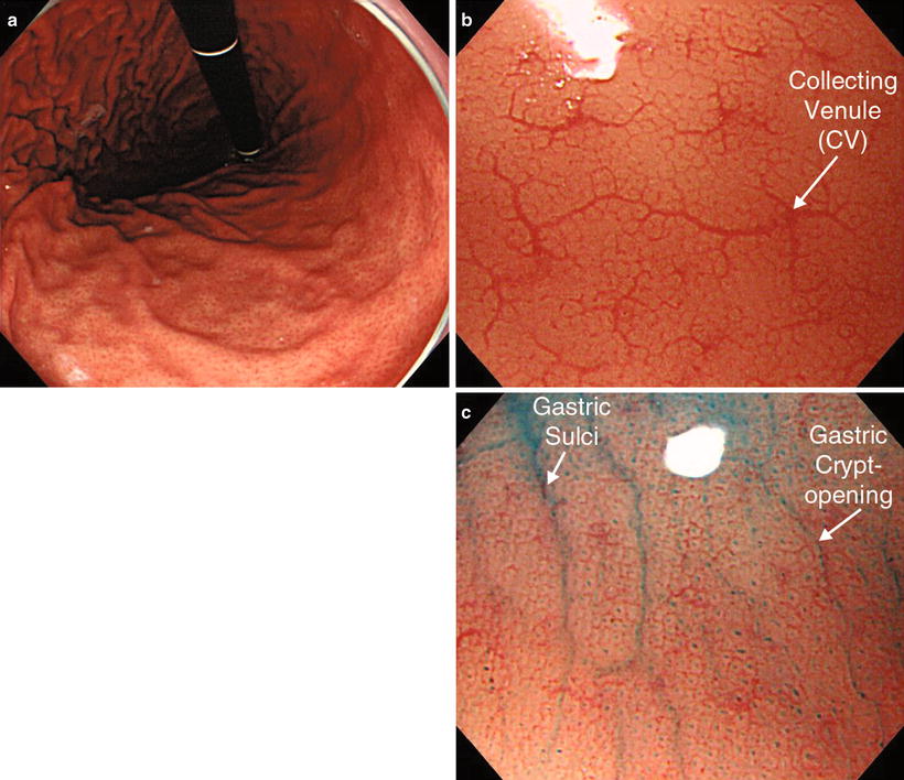
Fig. 4.1
Magnified endoscopic (ME) examination using white-light imaging (WLI) of normal gastric body (fundic gland region) (reprinted from [1] by permission of Dig Endosc). (a) Non-magnifying examination. (b) Magnifying examination. (c) Magnifying examination with indigo carmine dye spraying
4.2.2 Microsurface Structure (S)
Dye spraying makes the microsurface structure (S) more distinct. We see a regular oval crypt opening pattern, with round or oval crypt openings in an evenly spaced pattern, and at the same time, we can see the linear gastric sulci (Fig. 4.1c). The normal in vivo magnified endoscopic findings in the gastric body, including the capillary pattern, were first described by Yagi et al. [3, 4].
4.2.3 Pathological Confirmation
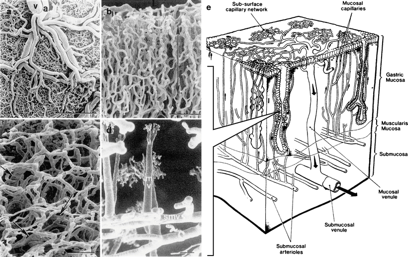
These magnified endoscopic findings have already been confirmed using scanning electron microscopy of vascular casts [5, 6]. In other words, as shown in Fig. 4.2, capillaries branch from submucosal arterioles, penetrating the muscularis mucosae. They are distributed from the base of the glands in the lamina propria towards the mucosal surface as they repeatedly anastomose with each other, surrounding the gastric glands. They form a regular honeycomb-like SECN, feeding into CVs in the subepithelial layer. The CVs travel obliquely downwards within the lamina propria to perfuse the submucosal venules.
 Basic Principles for the Interpretation of Magnified Endoscopic (ME) Findings: Vessels (V) Plus Surface (S) Classification System
The Proposed Vessels Plus Surface (VS) Classification System: Principles for Interpretation of Magnifying Endoscopy with Narrow-Band Imaging (M-NBI) Findings
Basic Principles for the Interpretation of Magnified Endoscopic (ME) Findings: Vessels (V) Plus Surface (S) Classification System
The Proposed Vessels Plus Surface (VS) Classification System: Principles for Interpretation of Magnifying Endoscopy with Narrow-Band Imaging (M-NBI) Findings
 Microanatomies as Visualized Using Magnifying Endoscopy with Narrow Band Imaging in the Stomach: Which Microanatomical Structures Can We Visualize in the Glandular Epithelium Using Narrow Band Imaging, and How Is This Achieved?
 Light Blue Crests (LBCs) and White Opaque Substance (WOS)
 Magnifying Endoscopy (ME) of the Stomach Targeting the Microvascular Architecture
 Analysis and Interpretation of Magnifying Endoscopy with Narrow Band Imaging (M-NBI) Findings in Gastric Epithelial Tumors (Early Gastric Cancer and Adenoma) Stratified for Paris Classification of Macroscopic Appearance


Fig. 4.2
Microvascular architecture of mucosa of the gastric body and fundus. (a–d) Scanning electron microscopic images of vascular casts. (a) Submucosal aspect (rat). Arterioles (a) and venules (v) branch in the submucosa, running in parallel. In the spaces between, we can see a mesh-like deep mucosal capillary network. Bar = 1 mm. (b) Vertical section of mucosa (rat). Capillaries run perpendicularly towards the luminal surface (arrow), repeatedly anastomosing on the way. Bar = 100 μm. (c) Image of capillaries from the luminal side (human). Polygonal capillaries line the crypt borders in a neat arrangement. Below the surface layer capillaries, we can see branches of the collecting venules (arrows). Bar = 100 μm. (d) Some collecting venules and capillaries (rat). The collecting venule (mv) is perfused by a subepithelial capillary (d) and is continuous with the submucosal venous plexus (smv). Bar = 100 μm. (e) Schematic diagram combining the findings from a–d (a, b and d reprinted from [4] by permission of the Journal of Anatomy, c reprinted from [5] by permission of Gastroenterology)
< div class='tao-gold-member'>
Only gold members can continue reading. Log In or Register to continue
Related posts:
Basic Principles for the Interpretation of Magnified Endoscopic (ME) Findings: Vessels (V) Plus Surface (S) Classification System
The Proposed Vessels Plus Surface (VS) Classification System: Principles for Interpretation of Magnifying Endoscopy with Narrow-Band Imaging (M-NBI) Findings
Microanatomies as Visualized Using Magnifying Endoscopy with Narrow Band Imaging in the Stomach: Which Microanatomical Structures Can We Visualize in the Glandular Epithelium Using Narrow Band Imaging, and How Is This Achieved?
Light Blue Crests (LBCs) and White Opaque Substance (WOS)
Magnifying Endoscopy (ME) of the Stomach Targeting the Microvascular Architecture
Analysis and Interpretation of Magnifying Endoscopy with Narrow Band Imaging (M-NBI) Findings in Gastric Epithelial Tumors (Early Gastric Cancer and Adenoma) Stratified for Paris Classification of Macroscopic Appearance
Stay updated, free articles. Join our Telegram channel
Full access? Get Clinical Tree
