Fig. 1
Schematic view of the hydraulic occluder used for induction of renal ischemia during MRI . 1 = indistensible extension tube, 2 = sutures, 3 = distensible silicone tube, 4 = renal vein, 5 = renal artery. A water-filled syringe, connected to the indistensible extension tube, is used to create a hydraulic pressure, which leads to an inflation of the distensible tube. This causes a compression of the renal artery and vein and restricts the blood flow. Adapted from ref. 10 with permission from the Public Library of Science
6.
Interferometric temperature measurement system (ACS-P4-N-62SC, Opsens, Quebec City, Canada), including a fiber-optical temperature probe (OTP-M, AccuSens, Opsens). If you use a GaAs crystal-based system instead be aware of the offset caused by the magnetic field (approx. 4.7 °C at 9.4 T).
2.3 Magnetic Resonance Imaging
Magnetic resonance imaging (MRI ) requires access to an ultra-high field MRI system including suitable accessories for the MR acquisition (radio frequency antennas), positioning, anesthesia, warming, and monitoring of physiological parameters, and trained personnel for operating the MRI system.
Due to the small size of rats in comparison with humans, a much higher spatial resolution is required to depict the kidney with adequate detail. This in turn demands a high signal-to-noise ratio (SNR), which must be achieved by use of tailored MR equipment.
1.
MR system: A dedicated small animal MR system with a magnetic field strength of 7 T and higher is recommended. Here we describe the use of a 9.4 T 20 cm bore system (BiospecTM 94/20, Bruker Biospin, Ettlingen, Germany) equipped with a gradient system with integrated shim set (B-GA12S2, Bruker Biospin; gradient amplitude 440 mT/m, max. slew rate 3440 T/m/s).
2.
Radio frequency (RF) coils: Use RF coils (antennas for RF transmission and reception) suitable for abdominal imaging, such as a transmit/receive rat body volume coil (72 mm inner diameter, quadrature; Bruker Biospin, Ettlingen, Germany) or preferably a transmit only rat body volume coil (72 mm inner diameter, linear; model T10325V3, Bruker Biospin) in combination with a receive only rat heart coil array (curved, 2 × 2 elements; model T12814V3, Bruker Biospin). Use of the latter coil setup is assumed here, as it allows for much higher spatial resolution due to its superior SNR when compared with the transmit/receive volume coil.
3.
Animal holder: An animal holder (here model T11739, Bruker Biospin) designed for the size of the animals and the geometry of the RF coils is provided by the MR system/RF coil manufacturer (see Note 1 ).
4.
Gases: O2, N2, and compressed air, as well as a gas-mixing system (FMI Föhr Medical Instruments GmbH, Seeheim-Ober Beerbach, Germany) to achieve required changes in the oxygen fraction of inspired gas mixture (FiO2). The following gas mixtures are required during the experiment: (i) for hypoxia —10 % O2/90 % N2; (ii) for hyperoxia —100 % O2; (iii) for normoxia—21 % O2 (air).
5.
Device for FiO2 monitoring in gas mixtures: for example Capnomac AGM-103 (Datex GE, Chalfont St Giles, UK).
6.
Device for warming of animal: Use a circulating warm-water-based heating system, consisting of a flexible rubber blanket with integrated tubing (part no. T10964, Bruker Biospin) connected to a conventional warm-water bath (SC100-A10, ThermoFisher, Dreieich, Germany). For alternative coil setups, water pipes may be built into the animal holder.
7.
Monitoring of physiological parameters: For monitoring of respiration and core body temperature throughout the entire MR experiment, use a small animal monitoring system (Model 1025, Small Animal Instruments, Inc., Stony Brook, NY, USA), including a rectal temperature probe and pneumatic pillow.
8.
Data analysis: Quantitative analysis of the data requires a personal computer and MATLAB software (R13 or higher; The Mathworks, Natick, MA, USA), ImageJ (Rasband, W.S., ImageJ, U.S. National Institutes of Health, Bethesda, Maryland, USA, http://imagej.nih.gov/ij/, 1997–2014), or a similar software development environment. Analysis steps described in the Subheading 3.7-3.13 can be performed manually by using the functions provided by the software development environment. Most of these steps benefit from (semi-)automation by creating software programs/macros—these steps are indicated by the computer symbol ( ).
).
 ).
).3 Methods
3.1 Preparation of MRI
1.
Start the ParaVision TM 5 software and—only before the very first experiment—create and store the following MR protocols.
(a)
Protocol_TriPilot (pilot scan): conventional FLASH pilot with seven slices in each direction (axial, coronal, sagittal).
(b)
Protocol_T2axl (axial pilot scan): RARE sequence, repetition time (TR) = 560 ms, effective echo time (TE) = 24 ms, RARE factor 4, averages = 4. Define as geometry an axial field of view (FOV) = 70 × 52 mm2, matrix size (MTX) = 172 × 128, eight slices with a thickness of 1.0 mm and distance of 2.2 mm, and an acquisition time of approximately a minute. Respiration trigger on (per phase step), flip-back on, fat saturation on.
(c)
Protocol_T2corsag (coronal/sagittal pilot scan): like Protocol_T2axl, but only one slice in coronal orientation.
(d)
Protocol_PRESSvoxel (shim voxel): conventional PRESS protocol, with a voxel size of = 9 × 12 × 22 mm3.
(e)
Protocol_MGE (T 2* mapping): multi-gradient echo (MGE) sequence, TR = 50 ms, echo times = 10, first echo = 1.43 ms, echo spacing 2.14 ms, averages = 4. Define as geometry a coronal oblique image slice with a FOV = (38.2 × 50.3) mm2, MTX = 169 × 113 zero-filled to 169 × 215, and a slice thickness of 1.4 mm. Respiration trigger on (per slice), fat saturation on.
(f)
Protocol_TOF (angiography): FLASH sequence, TR = 11 ms, TE = 3 ms, flip angle = 80°, spatial in-plane resolution of 200 × 268 μm2, with 15 slices of 1.0 mm thickness.
2.
Switch on the gradient amplifiers of the MR system, which will also power on the automatic animal positioning system AutoPac TM.
3.
Install the transmit/receive rat body volume coil in the magnet bore.
4.
Connect the animal holder to the animal positioning system (AutoPac TM).
5.
Install the rat heart coil array RF coil including its preamplifier on the animal bed.
6.
Attach the face mask unit (commercial or custom-made, as described in Note 1 ) to the animal holder and connect it to the inspiratory gas providing system (luer tubing).
7.
Place the flexible rubber mat of the warm-water-based heating system on top of the rat heart coil array and connect it to the warm-water circulation.
9.
Attach the rectal temperature probe and pneumatic pillow to the small animal monitoring system and place the probes on the animal bed, at the lower abdominal position of the rat.
3.2 Surgical Preparation
Surgery must be performed parallel to MRI preparation outside the MR scanner room (in a neighboring preparation room) for safety reasons.
1.
Anesthetize the animal by intraperitoneal injection of urethane (20 % solution, 6 mL/kg body mass) (see Note 4 ).
2.
After reaching the required depth of anesthesia (determined by specific physiological signs such as muscle relaxation degree, absence of the paw withdrawal reflex, absence of the swallowing reflex, whisker movements, etc.), carefully shave the coat in the abdominal area of the rat (hair clipper Aesculap Elektra II GH2, Aesculap AG, Tuttlingen, Germany).
3.
Place the rat in supine position on a warmed-up (39 °C) temperature-controlled operating table and fix the paws of the animal to the table by means of sticky tapes.
4.
Open the abdominal cavity by a midventral incision (4–5 cm). Carefully dissect both renal arteries from the surrounding tissues.
5.
6.
Place a fiber-optical temperature probe in close proximity to the kidney, in order to monitor the temperature of the kidney throughout the investigation.
7.
Mark the localization of the investigated kidney’s upper and lower pole on the skin of the abdomen. This is essential for optimal positioning of the rat in the MRI scanner (i.e., optimal position of the rat’s kidney relative to the MR coil).
8.
Fill the abdominal cavity with warm saline (37 °C). For replenishment of abdominal saline, a catheter must be placed in the abdominal cavity.
9.
Leave the tube/cable extensions of the occluder, of the catheter used for abdominal flushing, as well as the temperature probe, through the caudal cutting edge of the median abdominal incision.
10.
Close the abdominal cavity by continuous suture.
3.3 Transport, Mask, Positioning
2.
Position the rat supine on the MRI animal bed, while aligning the kidney (pen markings on skin) with the center of the RF surface coil array.
3.
Place a respiratory mask loosely around the muzzle of the spontaneously breathing rat. Open air supply to a rate of 1000 mL/min.
4.
Switch on the small animal monitoring system. Insert the rectal temperature probe after cleaning it with alcohol and dipping it into Vaseline™.
5.
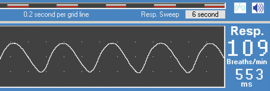
Place the pneumatic pillow on the abdomen and cover the animal with the warming blanket. Watch the respiration trace on the monitor of the small animal monitoring system and adjust pillow position until the respiratory motion is captured well (see Fig. 2) (see Note 8 ).


Fig. 2
Small animal monitoring system (SA Instruments, see Subheading 2). Setup of respiratory triggering for the MRI acquisition, with begin delay of 30 ms and maximum width of 280 ms for a typical respiratory rate of ~110 per minute. Horizontal white bars at top indicate trigger window
6.


Set the trigger options of the small animal monitoring system such that the trigger gate (indicated by white/red color horizontal bars parallel to the respiratory trace) opens for a maximum 280 ms (delay 30 ms) around the expiratory peak (see Fig. 2).
Stay updated, free articles. Join our Telegram channel

Full access? Get Clinical Tree


