Magnetic Resonance Imaging Acronyms and Abbreviations
To many radiologists, the world of magnetic resonance imaging (MRI) is like a secret society, consisting of a few radiologists, physicists, and equipment manufacturers who converse in their own incomprehensible language consisting of nonintuitive abbreviations and acronyms. It seems that the proliferation of acronyms and abbreviations has impeded rather than enhanced the dissemination of MR knowledge and created an air of intimidation and frustration surrounding abdominal and pelvic MRI.
We have found that MRI of the abdomen and pelvis becomes much less intimidating to our residents when we remind them that most of the available MR pulse sequences are designed to create one of two things—a T1-weighted image or a T2-weighted image (intermediate-weighted or proton density images are rarely used in abdominal and pelvic imaging). Furthermore, most pulse sequences generate these images using either a spin echo or gradient echo. Therefore, no matter what they are called, most abdominal and pelvic MR sequences are one of four basic types: spin echo T1-weighted, spin echo T2-weighted, gradient echo T1-weighted, or gradient echo T2-weighted (this latter sequence is more correctly referred to as T2*-weighted, because gradient refocusing fails to correct for magnetic field heterogeneities). As an additional modification, MR sequences may involve measuring a single echo per excitation (e.g., standard spin echo or gradient echo), multiple echoes per excitation (e.g., fast spin echo or echo planar), or all necessary echoes per excitation (single shot) (Fig. 4.23).
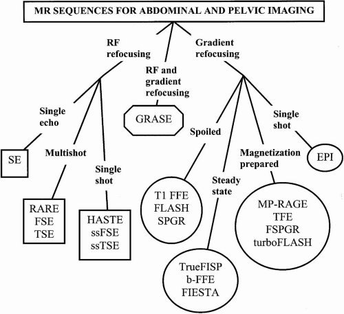 FIG. 4.23. Sequences for abdominal and pelvic MRI. This figure illustrates the differences between various MR sequences that are likely to be encountered in abdominal and pelvic MRI and the relationships between different acronyms and abbreviations. |
All pulse sequences are not available on all scanners. This is a source of endless frustration for radiologists desiring to apply sequences described in the literature to their own practice. Some sequences must be purchased (at a premium) from the scanner manufacturer. Others are only made available to beta test sites, or they require special hardware such as unique surface coils or high-performance gradients.
This section presents the more commonly encountered vendor-specific acronyms and abbreviations related to abdominal and pelvic MRI. Despite the large number of MR acronyms and abbreviations, certain generalizations can be made that may help one interpret a vendor-specific term. For example, G, GR, GE, or GRE usually refers to gradient echo. Field echo (FE) is another term for gradient echo. In general, the terms fast and turbo imply a rapid sequence that reduces scan time through a multishot technique. The letters IR in a term imply the use of an inversion recovery pre-pulse.
ASSET: Array spatial sensitivity encoding technique
See SENSE.
BALANCED FFE: Balanced fast field echo See FISP/trueFISP.
DRIVE: Driven equilibrium
Driven equilibrium techniques use a 90-degree radiofrequency (RF) pulse combined with a gradient refocusing pulse and a spoiling gradient to force magnetization from the transverse back into the longitudinal plane. DRIVE allows the TR to be reduced for echo train spin echo sequences by shortening the T1 relaxation time of fluid (which reduces the waiting period before the next excitation pulse). The shorter TR results in shorter imaging times without compromising the high signal intensity of fluids. DRIVE is primarily used for three-dimensional (3D) fast/turbo spin echo imaging.
EPI: Echo planar imaging
EPI acquires multiple gradient echoes per excitation. EPI can be performed as a single-shot or multishot technique but requires high-performance gradients (not available on some systems). Several EPI acquisition schemes exist, all of which allow for extremely rapid imaging. However, EPI may be limited by artifacts (e.g., susceptibility). EPI is currently seldom used for abdominal and pelvic imaging, although new abdominal and pelvic applications are being developed.
FIESTA: Fast imaging employing steady-state acquisition
See FISP/TrueFISP
FISP/TrueFISP: Fast imaging with steady-state precession (Fig. 4.24)
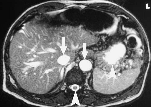 FIG. 4.24. Axial image of upper abdomen obtained with balanced-fast field echo sequence (b-FFE, FIESTA, trueFISP). Note bright vessels (arrows) and fluid (arrowheads). |
A steady-state gradient echo sequence, FISP samples the free induction decay signal. No spoiling is applied, allowing both longitudinal and transverse components of magnetization to persist in a steady state. This original sequence is seldom used in abdominal and pelvic imaging. Similar sequences are GRASS, FAST, and FFE. In trueFISP imaging, the free induction decay signal and spin echo/stimulated echo signal are collected simultaneously, resulting in a predominately T2/T1-weighted image. Fluid and intravascular blood appear bright and tissue interfaces are sharp with this sequence. Flow artifacts seen with HASTE may be eliminated with trueFISP. These sequences are useful for imaging fluid-filled structures, such as the biliary tract, pancreatic duct, and urinary collecting system, as well as for vascular and fetal imaging. TrueFISP is similar to balanced FFE and FIESTA.
FLASH: Fast low-angle shot (Fig. 4.25).
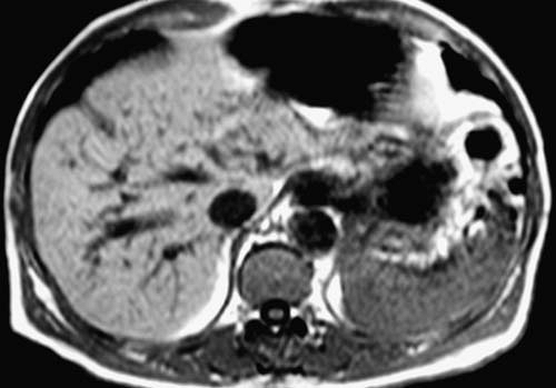 FIG. 4.25. Axial image of upper abdomen obtained with T1-weighted spoiled gradient echo sequence (FFE, FLASH, SPGR).
Related posts:Stay updated, free articles. Join our Telegram channel
Full access? Get Clinical Tree
 Get Clinical Tree app for offline access

|


