Lymphoproliferative Disorders of the Gastrointestinal Tract


Lymphoproliferative Disorders of the Gastrointestinal Tract


OVERVIEW


Introduction

The gastrointestinal (GI) tract contains the largest aggregate of lymphoid tissue aside from lymph nodes, and it is the most frequent site of extranodal lymphoma.1, 2 Surprisingly, the most common site of lymphoma in the GI tract is the stomach, which normally has only rudimentarily organized lymphoid tissue. Advances in molecular genetics have led to a better understanding of the pathogenesis of GI lymphoproliferative disorders and have had a great impact on how we approach these lesions in clinical practice today.

Understanding the organization of normal lymphoid tissue in the gut is essential to understanding its lymphoproliferative disorders. A detailed description of GI lymphoid tissue and its functions is provided in Chapter 5, and only a brief discussion is presented here.3 Aside from the mesenteric lymph nodes, the GI lymphoid tissue is represented by organized mucosa-associated lymphoid tissue (MALT), intraepithelial lymphocytes (IELs), and lamina propria lymphoid cells. Of these, MALT and IELs are unique in many ways.

MALT in the GI tract refers to unencapsulated lymphoid aggregates associated with specialized epithelium, best represented by Peyer’s patches in the terminal ileum and lymphoid tissue of the appendix. However, they are widely distributed throughout the GI tract and vary in size and numbers considerably, not only among different individuals but also in the same individual over time. Similar to lymph nodes, these aggregates have organized B- and T-cell zones along with many accessory cells. The B-cell zone consists of lymphoid aggregates, which may develop germinal centers when stimulated, each of which is surrounded by an outer mantle and an outermost marginal zone similar to lymph nodes. The marginal zone is a broad area populated by small- to intermediate-sized cells that morphologically resemble germinal center cells (centrocyte-like cells [CLC]) or monocytes (monocytoid B cells). The marginal zone cells extend toward the epithelium and can be seen entering the overlying epithelium constituting “lymphoepithelium,” which is the defining feature of MALT. These lymphocytes are IgM-positive and IgD-negative or weakly positive, as compared to mantle zone cells that are positive for both IgM and IgD. The follicles are flanked on the lateral as well as the basal aspects by a T-cell area similar to the paracortical T-cell zone of the lymph nodes.

The lamina propria throughout the gut is populated by a mixture of plasma cells, lymphocytes (B and T cells), mast cells, eosinophils, and macrophages. The composition and the density of this population vary between different GI segments among individuals and even within an individual over a period of time. The plasma cells secrete predominantly IgA but also IgM, IgG, and IgE. The CD4:CD8 ratio of the T-cell population is approximately 4:1. About 50% of these cells express CD103, which represents integrin α4β7 that plays a role in the adhesion of these cells to the epithelium and lymphocyte homing to the gut.

IELs away from MALT areas are largely T cells of a heterogeneous phenotype. They are widely present throughout the GI tract, including the squamous mucosa. The predominant phenotype is the cytotoxic T-cell expressing αβ T-cell receptors (TCRs), which are CD3+, CD8+, CD103+, CD4−, and CD5−. A smaller population (10%-15%) consists of T cells expressing γδ TCRs that are negative for both CD4 and CD8. A third population of CD56+ IELs is also recognized that are virtually undetectable in normal mucosa. They T-cytotoxic and natural killer (NK) properties.

Studies, largely from experimental animals, show that lymphocytes from MALT have receptors that recognize specific cellular adhesion molecules on high endothelial venules that regulate their circulation and homing. This applies to both B and T cells. The MALT obviously functions as the host’s immune response to various luminal organisms and allergens. IELs are known to increase in response to infections or food-induced enteropathies and are also likely to be an important component of the host’s immune response; however, their specific function is poorly understood, although as predominantly CD8+ cells they likely downregulate any immune response.


Incidence

The GI tract accounts for about 50% of all primary extranodal lymphomas. In addition, GI involvement occurs in up to 50% of patients with disseminated nodal lymphomas or lymphocytic leukemias, in autopsy studies.8, 9 Nevertheless, in the United States, GI lymphomas are still relatively uncommon compared to other GI tumors, primarily adenocarcinomas. Thus, although more than half of all primary and secondary GI lymphomas in the United States are of gastric origin, they represent only about 5% of all gastric tumors.

It should also be noted that there is a marked geographic variation in the types and the site predilection of GI lymphomas (see Table 4-2). In the United States and Europe, the most frequent site of GI lymphomatous involvement is stomach (50%), followed by the small intestine (37%) (primarily the ileum and the ileocecal region) and anorectal region (3%).2, 10, 11, 12, 13 Involvement of the remainder of the intestinal tract, such as duodenum and mesentery, is uncommon.14 In contrast, in the Middle East and South Africa, the incidence of intestinal lymphomas is much more common than that of gastric lymphomas, ranging from 35% to 75% of all GI lymphomas, due to a relatively higher incidence of Mediterranean lymphoma and α-chain disease.15 Interestingly, the duodenal lymphomas occur most frequently around the ampulla of Vater and consist primarily of follicular lymphomas, which are exceedingly rare in other portions of the GI tract.16


Pathogenesis of GI Lymphoma

The predisposing conditions for GI lymphoma are varied and include infections such as Helicobacter pylori (H. pylori) celiac disease, diffuse and nodular lymphoid hyperplasia (which themselves are likely secondary), immunodeficiency disorders (primary or acquired), and occasionally familial tendency. Lymphoma has also been described in association with ulcerative colitis, Crohn’s disease, and epithelial neoplasms.

Details of the pathogenesis of the common GI lymphomas are unraveling a number of interesting associations.3 While the pathogenesis of the nodal lymphomas remains unclear, primary GI lymphomas have led the way in understanding the “genesis” of lymphoma. The key theme of chronic antigenic stimulation (infectious or noninfectious) and immune dysregulation leading to benign proliferations that may eventually lead to a malignant disease is exemplified by many primary GI lymphomas. This process is in a way similar to the “hyperplasia-adenoma-carcinoma” sequence of epithelial neoplasms, although it is currently difficult to define the concept of “prelym-phoma” or “lymphocytic dysplasia.” Understanding the pathogenic mechanisms has had a great impact not only on the understanding of these disorders but on their management as well. A leading example of this is H. pylori-related gastric MALT lymphoma, where treatment of H. pylori results in disappearance of the lymphoma. As our understanding of H. pylori-induced gastric MALT lymphoma continues to evolve, the search for infectious etiologies of other types of lymphomas has already begun to show promising results. Infectious agents that have been implicated in the etiopathogenesis of other lymphomas include Campylobacter jejuni, Clamydia psittaci, Borrelia burgdoferi, Epstein Barr virus (EBV), and hepatitis B
and C viruses.17, 18, 19, 20 Of these, EBV has already been a subject of interest for many years.


Classification of GI Lymphomas

The accurate classification of GI lymphomas is important for identifying specific disease entities and also for determining treatment protocols and assessing prognostic factors. The literature on GI lymphoma suffers from the use of nonuniform classification systems, making it difficult to compare results.

Until fairly recently, the major lymphoma classifications used were the Working Formulation, Rappaport’s classification, WHO modification of the Working Formulation, and Kiel classification, all of which were based primarily on morphologic and immunophenotypic features.21, 22, 23, 24 The problem with the Working Formulation was that the classification was originally developed for the nodal lymphomas, and it was found that many extranodal lymphomas were not variants of nodal lymphomas but were histologically and clinically distinctive. For example, MALT lymphoma, IPSID, and enteropathy-associated T-cell lymphoma (EATL) have histologic features quite distinct from the nodal lymphomas. The Kiel classification was also morphologic based and divided lymphomas primarily into two broad categories, namely, B and T cells, and then classified them on the basis of cytologic morphology and grading. Although the grading frequently predicted the clinical outcome, lymphomas like the mantle cell lymphomas that were morphologically considered low grade are actually aggressive. Other lymphomas, such as the anaplastic large-cell lymphomas, that were considered high grade and to have an aggressive natural history show excellent response to chemotherapy with prolonged disease-free survival.25


Table 4-1 Tumor Site of Gastrointestinal Lymphomas


TUMOR SITE


TUMOR TYPE


USUAL


LESS FREQUENT SITES


B-CELL TYPE


GUT TYPE


MALT lymphoma


Stomach


Esophagus, small intestine, and large intestine


IPSID and α-chain disease (Mediterranean lymphoma)


Small intestine


Stomach, colon


NODAL TYPE (primary or secondary)


Mantle-cell lymphoma


Ileocecal region and stomach


Follicular center cell lymphoma


Ileocecal region


Colon and stomach


Diffuse large B- cell lymphoma


Small intestine


Colon and stomach


Burkitt’s and Burkitt’s-like type


Ileocecal region


Throughout the gastrointestine


AIDS-associated lymphoma


Stomach


Colon, ileum, esophagus, and duodenum


Miscellaneous B-cell TYPE


PTLD


Throughout the GI tract


Plasmacytoma


Stomach and small intestine


T-CELL TYPE


GUT TYPE


Enteropathy associated T-cell (EATL)


Jejunum


Remainder small intestine and stomach


Mesenteric lymph nodes


NODAL TYPE (Primary or secondary)


Nonenteropathy associated lymphoma


Throughout the GI tract


Angiocentric lymphoproliferative lesion


Throughout the GI tract and mesentery


Mycosis fungoides


Throughout the GI tract and mesentery


MALT, mucosa-associated lymphoid tissue; IPSID, immunoproliferative small intestinal disease; PTLD, posttransplant and other iatrogenic lymphoproliferative disorders.


To address these issues, in 1994 a new lymphoma classification was introduced by the International-American Lymphoma Study Group, labeled as the Revised European American Lymphoma (REAL) classification.26 This classification subdivided lymphomas into distinctive entities, based on multiple parameters, including clinical presentation, site of the primary lesion, natural history and clinical course, morphologic appearance, and immunophenotypic and genetic factors. The site of the primary lesion
is important for most extranodal tumors, including many GI lymphomas. This REAL classification was upgraded in 1999 and has now been adopted by the WHO classification.27, 28, 29 While the lymphoma classifications will continue to evolve, disease-specific rather than morphology-centered classification is likely to remain a central theme.

The classification of GI lymphomas suggested here is based on the REAL and WHO classifications (Table 4-1). A fair number of distinctive lymphomas occur in the GI tract, although the frequency of the specific types varies greatly from one site to another (Table 4-2).

The vast majority (85%) of GI lymphomas are of B-cell origin, of which most are MALT lymphomas. T-cell lymphomas are rare and occur most often in the small intestine accounting for 34% of cases in one study. The latter are often associated with celiac disease. Since the advent of AIDS and organ transplantation, several new types of lymphomas have been recognized that can involve the GI tract. Only a handful of patients with Hodgkin’s disease and other tumors, such as solitary plasmacytoma, large-cell anaplastic lymphoma, and true histiocytic neoplasms, have been described in the GI tract. The GI tract is also commonly involved secondarily in advanced stages of nodal lymphomas and leukemia. Lastly, a number of lymphomas are impossible to subclassify beyond their lymphoid derivation and are designated as unclassified.


Table 4-2 Classification of GI Lymphomas


B CELL


T CELL


MISCELLANEOUS


GUT TYPE


  1. MALToma (extranodal marginal zone B-cell lymphoma)


  2. IPSID and α-chain disease (Mediterranean lymphoma)


    1. Stage A


    2. Stage B


    3. Stage C/DLBCL


NODAL TYPE (PRIMARY OR SECONDARY)


  1. Follicular lymphoma


  2. Mantle-cell lymphoma


  3. Burkitt’s and Burkitt’s-like lymphoma


  4. DLBCL


  5. SLL


  6. Lymphoplasmacytic lympoma


  7. Anaplastic large-cell lymphoma B-cell type


  8. Plasma cell myeloma/plasmacytoma


  9. Plasmablastic lymphoma


  10. T-cell rich B-cell lymphoma


  11. Angiocentric lymphoproliferative lesion (lymphomatoid granulomatosis)


MISCELLANEOUS B-CELL LYMPHOMAS


  1. Primary immunodeficiency-associated lymphoma


  2. HIV-associated lymphoma


  3. PTLD


    1. Plasma cell hyperplasia (polyclonal)


    2. Polymorphous—usually monoclonal


    3. Lymphoma/myeloma (plasmacytoma)


  4. Other iatrogenic and mediation-associated lymphomas


GUT TYPE


Enteropathy associated T-cell lymphoma (EATL)


Small intestinal CD4 positive lymhoma


NODAL TYPE (PRIMARY OR SECONDARY)


  1. Peripheral T-cell lymphoma, not otherwise categorized


  2. Mycosis fungoides/Sezary’s syndrome


  3. Angioimmunoblastic T-cell lymphoma


  4. Anaplastic large-cell lymphoma T-cell type


MISCELLANEOUS T-CELL LYMPHOMAS


PTLD


Monomorphic T/NK-cell type


  1. Hodgkin’s disease


  2. PTLD


    Hodgkin’s type


  3. Histiocytic lymphoma (Histiocytic sarcoma)


  4. Langerhans cell histiocytosis


  5. Mastocytosis


  6. Unclassified lymphoma


  7. Leukemic involvement of the GI tract. (Granulocytic sarcoma)


  8. Composite lymphoma


    Mixed type lymphoma


    Carcinoma/lymphoma


MALT, mucosa-associated lymphoid tissue; IPSID, immunoproliferative small intestinal disease; PTLD, posttransplant and other iatrogenic lymphoproliferative disorders


Role of Molecular Diagnosis in GI Tract Lymphomas

Molecular diagnostics has made significant advances in the recent years, and the utility of various techniques has been well established.31, 32 Diagnosis and proper classification of lymphoma in current practice is based on a multidisciplinary approach, in which clinical features, morphology, immunopheno-type, and cytogenetic and molecular characteristics are all important to varying extent in a given case. In practice, the diagnosis of lymphoma can be established with morphology and immunohistochemical analysis in most cases, while in some cases additional evidence of monoclonality may be essential in establishing a definitive diagnosis. In addition to clonality assessment, molecular techniques also help in some cases to establish the cell lineage, correctly classify the lymphoma, differentiate Hodgkin’s from non-Hodgkin’s lymphoma, and detect minimal residual disease.


Table 4-3 Modified Ann-Arbor Staging System for Primary Gastrointestinal Lymphomas


IE


Localized involvement without lymph node metastases


IE-1


Early lymphoma. Tumor confined to the mucosa and submucosa


IE-2


Lymphoma extends beyond the submucosa


IIE


Lymphoma of any depth of infiltration with lymph node metastases


IIE-1


Lymphoma with contiguous regional node metastases


IIE-2


Lymphoma with lymph node metastases beyond contiguous regional nodes


IIIE


Localized lymphomatous involvement with lymph nodes on both sides of the diaphragm (from a practical point almost never applies to the stomach)


IVE


GI lymphoma with or without associated lymph node metastases with diffuse lymphomatous dissemination beyond the GI tract


The suffix E denotes extranodal.


The targets for molecular diagnosis of lymphoma in routine practice are (a) rearrangement of immunoglobulin (Ig) and TCR genes, (b) specific chromosomal translocations, and (c) viral infection (e.g., EBV) in certain situations.32

The commonly used molecular techniques in clinical practice include (a) southern blot analysis, (b) polymerase chain reaction (PCR) and reverse transcription PCR (RT-PCR), and (c) fluorescence in situ hybridization (FISH) or RNA in situ hybridization. Other evolving technologies that may find a significant role in the clinical practice in future include spectral karyotyping and gene expression profiling by cDNA microar-ray. Southern blot analysis remains the gold standard in clonality testing. However, it requires fresh tissue for obtaining large amounts of high-quality DNA, is labor-intensive, requires longer time, uses radioactive materials in the detection system, and has a low analytical sensitivity. In most labs, PCR-based assays have replaced southern blot assays for molecular diagnostics and are the preferred first-line approach. They have the following advantages: they require less tissue, they can use DNA or RNA as templates, DNA quality is less critical allowing fixed and archival material to be used, they have rapid turnaround time and superior sensitivity, and they do not require radioactive detection systems and can be automated. The electrophoretic systems that allow discrimination of PCR products have also evolved over time, and the newer systems, for example, capillary electrophoresis, have higher resolution allowing greater sensitivity of the assays. While this is extremely helpful in detection of minimal residual disease where neoplastic cells are expected to be scanty, they carry a disadvantage of identifying small clonal populations in benign reactive
conditions. Using sensitive techniques, about 10% to 15% cases of H. pylori-related chronic gastritis without any histologic evidence of lymphoma may show clonal B-cell populations. The choice of the molecular technique in a given situation depends upon the clinical setting, target to be assayed and the test availability. Further discussion of the technical aspects of molecular tests and their limitations are beyond the scope of this book.

The key role of molecular testing lies in the demonstration of monoclonality in difficult cases. The detection of Ig heavy chain gene rearrangement is the most commonly used assay for establishing B-cell clonality in most labs, although assays for Ig light chains are also available. The qualitative sensitivities of these assays vary widely from <50% to virtually 100% and depend upon assay design, primer selection, detection system, and case type. False-negative results are more likely to occur in follicular lymphomas, marginal zone lymphomas, and DLBCLs largely due to V-region somatic hypermutations, which affect primer annealing in complementary-determining region 3 assays.32 For assessment of T-cell clonality, the TCRγ gene is preferentially used in most labs due to simplicity of its genomic structure and requirement of relatively few Vγ and Jγ primers to detect all possible rearrangements.32 TCRγ rearranges fairly early in both αβ+ and γδ+ T cells and provides a suitable molecular target irrespective of the nature of the ultimate surface receptor expression. TCRβ gene rearrangement assays are also available but are less frequently used in practice.

Detection of specific lymphoma-associated translocations forms an important component of molecular tests and is used for diagnostic purposes, proper classification, detection of minimal residual disease, and follow-up. The translocations not only provide supportive evidence for clonal expansion of lymphocytes but also point toward specific pathogenic events in the lymphoma development. They may be detected by a variety of techniques including southern blot, DNA-PCR, RT-PCR, conventional cytogenetics, and FISH. The most commonly assessed translocation in this regard are t(14;18)(q32;q21) for follicular lymphoma, t(11;14)(q12;q32) for mantle lymphoma, and t(2;5)(q23;351) for anaplastic large-cell lymphoma.

Viral infections have been implicated in the development of many lymphomas, of which EBV is the most important from an etiologic as well as diagnostic stand point. The Demonstration of EBV is important in posttransplant lymphoproliferative disease and primary effusion lymphoma among many other conditions. EBV can be demonstrated in a number of ways using immunohistochemical, southern blot and PCR-based techniques, although the method of choice is in situ hybridization for EBER.32 This is relatively inexpensive and can be easily applied to paraffin sections with high sensitivity and specificity.

Among evolving newer technologies from a diagnostic clinical application stand point, gene expression profiling using cDNA microarray technology remains the most promising.32 The technique allows for genomic-scale gene expression profiling of tumors. It offers opportunity for refining existing lymphoma classification, provides data regarding expression of novel markers, and helps in development of targeted therapies and prognostication. cDNA chips are commercially available and can also be custom designed. The current technique requires undegraded mRNA from the tissue samples, and it is of utmost importance that laboratories harvest fresh tissue for any future use in this regard.

It cannot be overstated that clonality does not equate to malignancy, nor does its absence exclude malignancy. For example, a number of clonal but benign lymphocytic proliferations have been described at other sites, such as benign monoclonal gammopathy, cutaneous lymphoid hyperplasia, drug-induced dermatoses, myoepithelial sialadenitis, and idiopathic cold hemagglutinin disease, all of which have an increased but not absolute risk of malignant transformation.32 In many cases that eventually transform to malignancy, clonal identity can be shown between the clone detected at the initial reactive stage and the subsequent malignancy.33, 34 This would suggest that monoclonality might be an intermediate stage in the evolution of lymphoma, requiring additional steps before malignant transformation, very similar to the multistep carcinogenesis of epithelial tumors. It should also be recognized that well-defined cases of lymphoma fail to show IgH rearrangement by PCR in about 10% to 30% of cases.32 Possible reasons include poor DNA preservation, chromosomal abnormalities involving the immunoglobulin locus, or poor primer binding associated with ongoing mutations within the gene.

Clonal B-cell proliferations have also been detected in cases of follicular chronic gastritis, the rates varying greatly among studies.33, 35, 36 It has been shown that the clones fluctuate and may disappear or evolve over a period of time.37 In a prospective study of chronic gastritis with monoclonality and without any histologic suspicion of lymphoma, it has been shown that some cases evolve over time into definite lymphomas while some do not.37, 38 PCR performed retrospectively on prior benign biopsies in cases that eventually developed lymphoma has shown identical clonal B-cell populations in the absence of histologic evidence of lymphoma.33 While more studies are needed to clearly define the clinical significance of monoclonality in this setting and its long-term
outcome, in routine practice, presence of clonal B-cell proliferation in absence of histologic evidence of lymphoma cannot be totally disregarded.39

False-negative and false-positive results have been recognized in the literature with each molecular technique in use, and the rates tend to vary with techniques and among laboratories.32 The sensitivity and the specificity of currently used various molecular techniques in experienced laboratories generally exceeds 90%. However, tremendous interlaboratory variability exists and there is a lack of standardization of techniques. For the practicing pathologist it is important to realize the utility and limitations of each technique and know the sensitivity and the specificity of the assays used. It cannot be overemphasized that diagnosis of lymphoma should never be made in isolation entirely based on molecular assays, but should be interpreted in light of clinical context, histology, and other laboratory data.


Workup of Lymphoproliferative Disorders of the GI Tract

First, if the endoscopist suspects he or she is dealing with a case of GI lymphoma, the pathology lab should be alerted that the tissue is coming so it can be processed immediately, and some of the biopsies should be placed in saline or culture medium for flow cytometry and some in formalin. Many institutions have a “lymphoma protocol” that caters to all of these needs. In practice the pathologic diagnosis of lymphoma is made under two circumstances: (a) in clinically unsuspected cases where examination of the biopsy or resection specimen is the first indication of lymphoma or (b) lymphoma is either known or suspected prior to the biopsy or resection. In the first circumstance, there is often no choice. However, in the second situation care must be taken to appropriately triage tissues for lymphoma workup. Most of the common antibodies for immunohistochemical analysis needed for the diagnostic workup of lymphoproliferative disorders, gene rearrangement studies, in situ hybridization, and FISH for molecular genetic analysis can be performed on formalin-fixed paraffin-embedded tissues. Fresh tissues in appropriate media are needed for flow cytometry, conventional cytogenetics, and tissue cultures. Need for these additional studies is based on the complexity of a case and the academic and research interests at an institution.

Flow cytometry is an invaluable adjunct to diagnostic workup of lymphoproliferative disorders as it provides superior immunophenotyping opportunities and clonality assays based on unique immunophenotype and light chain restriction and provides very fast results. Flow cytometry is of somewhat limited use in the workup of T-cell lymphomas and more so for Hodgkin’s lymphomas. Nucleic acid extraction and PCR-based assays while often performed on paraffin-embedded tissues provide better yield with frozen tissue samples. Touch/imprint smears made from fresh tissues often provide better cytologic details while compromising architectural pattern recognition. They are invaluable not only in the immediate assessment of tissues with a suspected diagnosis of a lymphoproliferative disorder often for intraoperative consultation but could also help in deciding proper tissue triage for diagnostic workup. Based on the resources available and the needs, each laboratory must design a lymphoma protocol to be applied to suspected cases of lymphoma. In many laboratories, the tissues are initially handled by individuals in training or technicians, and the opportunity to adequately work up complex cases is inadvertently lost in some cases. It is of utmost importance to have a strong suspicion based on the clinical circumstances to appropriately triage tissues for a lymphoma workup. Pathologists, especially trainees, should not hesitate to consult the seniors or contact their clinical counterparts whenever the need arises. Often frozen sections and touch preparations prepared just to properly triage tissue specimens pay rich dividends.


LYMPHOID HYPERPLASIA OF THE GASTROINTESTINAL TRACT

Lymphoid hyperplasia occurs in virtually all parts of the GI tract. Its frequency is difficult to assess, as it is often physiologic, asymptomatic, and seldom biopsied.40 Diagnostic criteria to differentiate it from normal are lacking so the diagnosis of lymphoid hyperplasia is entirely subjective. In general, lymphoid nodules in a quiescent gut are not visible to naked eye, other than those in the terminal ileum. Visibility on naked eye examination or endoscopy that roughly corresponds to nodules >0.2 mm is an indirect and arbitrary criteria of hyperplastic lymphoid follicles. The situation is more complex when dealing with lymphoid hyperplasia at sites that normally contain abundant lymphoid tissues like terminal ileum and appendix. Unlike appendix, terminal ileal mucosa is accessible to visual examination to both gastroenterologists and pathologists, and everyone intuitively develops a sense of what is normal and what is not especially once normal Peyer’s patches can be identified. Regarding the appendix, its size is so variable that it cannot even be used for defining lymphoid hyperplasia.

There are several situations where lymphoid tissue may be recognized clinically including Helicobacter gastritis especially in children; in the duodenum
where it is a cause of nodularity; when appendiceal lymphoid tissue can be seen during endoscopic screening of mucosa at the appendiceal orifice and is biopsied as a “mucosal polyp”; in resected appendices, mostly for acute appendicitis, when obliteration of its lumen could be used as indirect evidence of lymphoid hyperplasia; in children in whom it may form the apex of an intussusception, often associated with adenovirus infection, and in diversion colitis/proctitis when the lymphoid hyperplasia can be seen grossly. Histologically, it is even more difficult to define lymphoid hyperplasia. The presence of lymphoid follicles with large secondary germinal center often correlates with grossly enlarged lymphoid nodules. The challenge is often to ensure that there are plasma cells present and that this is not a manifestation of common variable immunodeficiency (CVID) disease, and, if it is, that organisms such as Giardia are not present.

Lymphoid hyperplasia can be either localized or diffuse and can be assigned to one of five distinct clin-icopathologic groups:

1. Localized lymphoid hyperplasia of the stomach: gastric lymphoid hyperplasia.

2. Localized lymphoid hyperplasia of the small intestine (excluding terminal ileum).

3. Localized nodular lymphoid hyperplasia of the terminal ileum and appendix.

4. Localized lymphoid hyperplasia of the rectum (benign lymphoid polyp, rectal tonsil).

5. Diffuse nodular lymphoid hyperplasia.

On rare occasions lymphoid hyperplasias have also been described in unusual locations such as esophagus and colon.41, 42, 43, 44

Familiarity with various forms of GI lymphoid hyperplasia is important, particularly for its distinction from malignant lymphoma of the GI tract. Lymphoid hyperplasia may also occur as a secondary component in a number of disorders, which include nonerosive gastritis, immunodeficiency disorders, Crohn’s disease, ulcerative colitis, and diversion colitis, and may sometimes cause difficulty in diagnosis. However, in primary lymphoid hyperplasia, the proliferation of lymphoid tissue is the predominant or the only pathologic feature, which is responsible for producing topographic and structural changes.


Localized Lymphoid Hyperplasia of the Stomach—Gastric Lymphoid Hyperplasia (Pseudolymphoma)

The evolution of this entity and the term “pseudolymphoma” is very interesting. This concept was first suggested by Smith and Helwig in 1958.45 After reviewing all their cases of gastric lymphoma, they suggested that many cases actually represented benign reactive hyperplasia and that gastric lymphoma was overdiagnosed. This was based on the reactive-looking histology and excellent prognosis of these cases. The term pseudolymphoma was coined by Jacobs in 1963 for such lesions.46 Subsequently, the term gastric pseudolymphoma was widely used and understandably so.47, 48, 49, 50 After MALT lymphomas of the stomach were described by Isaacson in 1983, gastric pseudolymphomas became uncommon.51 In 1997 Abbondanzo and Sobin reviewed all their cases of gastric “pseudolymphoma,” and based on histology and immunostains they concluded that 77 of the 93 cases diagnosed as pseudolymphoma represented true lymphomas (51 MALT lymphomas, 10 mantle cell lymphomas, 9 DLBCLs, and 7 follicular lymphomas).52 Five cases were considered as atypical lymphoid infiltrate and only five were considered truly benign chronic follicular gastritis. Gene rearrangement and cytogenetic studies were not performed in this study and the follow-up was limited. The authors concluded that the majority of pseudolymphoma cases represent true lymphomas, and the term should be abandoned. Indeed, cases that represent extreme hyperplasia of gastric lymphoid tissue in current practice are extremely rare, and search of current literature fails to reveal any such convincing cases. The term gastric lymphoid hyperplasia, if used at all, should be confined to cases of chronic nonspecific gastric inflammation, in which the proliferation of lymphoid tissue presents as a tumorous mass, and the possibility of a lymphoma, especially of the MALT-type, has been excluded.

However, a diffuse fine nodularity is most commonly associated with H. pylori and tends to regress following its eradication.48, 53, 54 Patients usually present with symptoms suggestive of peptic ulcer disease or chronic gastritis, although occasionally they have gastric outlet obstruction.55

Pathology. Morphologically, these lesions consist of either fine gastric mucosal nodularity, primarily in children, or small nodular or microscopic lesions in adults.56 In these cases, the lymphoid infiltrate, often with germinal centers, produces mucosal thickening, which may be accompanied by either an ulcer with raised margins or a localized plaque-like thickening of the rugal folds without a recognizable ulcer (Fig. 4-1).

The lesions are usually solitary and small.

Mucosal ulceration, when present, is superficial. In most instances, these lesions seem to represent less advanced examples of gastric lymphoid hyperplasia in current practice, because of early recognition and treatment of H. pylori infection.

The typical gastric lymphoid hyperplasia is associated with a dense polymorphous inflammatory infiltrate,
admixed with lymphoid follicles with germinal centers and fibrosis (Fig. 4-2). Since reactive lymphoid follicles may also be found within gastric lymphomas, one cannot rely on these follicles for distinguishing lymphoid hyperplasia from lymphoma. Clumps of IELs mimicking lymphoepithelial lesions of MALT lymphomas may also occasionally be found, but they differ from the latter in that they consist of mature lymphocytes, mostly T cells, usually have less than three cells, and do not cause epithelial destruction or degeneration.


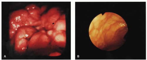
Figure 4-1. Endoscopic appearances of gastric lymphoid hyperplasia. A: Central ulceration surrounded by severe mucosal nodularity. B: Plaque-like thickening of rugal folds without ulceration.


Angiofollicular Hyperplasia (Lymphoid Hyperplasia with “Castleman-like” Features)

On very rare occasions, these variants of lymphoid hyperplasia have been reported in the stomach and small intestine.47, 57 Angiofollicular lymphoid hyperplasia of the GI tract is morphologically similar to its nodal counterpart, showing a lymphoid infiltrate with numerous lymphoid follicles containing hyalinized germinal centers and vascular structures.47 Unlike the usual gastric lymphoid hyperplasias, these lesions are not associated with ulceration or fibrosis. It is unclear if they have any significant clinical implication or just represent a histologic variation of reactive lymphoid hyperplasia. Its relationship to HIV infection has not been studied to date.


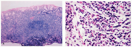
Figure 4-2. Endoscopic biopsy specimen of gastric lymphoid hyperplasia. A: Low-power view showing a dense mucosal inflammatory infiltrate. B: Detail of part (A) showing a polymorphous infiltrate composed of mature lymphocytes, plasma cells, eosinophils, and histiocytes.

Immunohistochemical features and molecular genetics. Immunohistochemistry of the lymphoid infiltrate is expected to show a typical polyclonal pattern of the immunoglobulin light chains. (In addition, there is a marked T-cell reaction occurring in a perivascular location in the mucosa and in submucosal nodules.58) Sometimes, it may be extremely difficult to differentiate between a lymphoid hyperplasia and a low-grade lymphoma on a small biopsy specimen, especially, in suboptimally fixed or distorted specimens. In such a situation, repeat biopsy with optimum fixation, immunohistochemistry, and gene rearrangement studies could be helpful (see also section on
differential diagnosis of MALT lymphoma).59 Presence of a clonally rearranged Ig gene detected by PCR in this setting is likely to represent a lymphoma, and the histology needs to be interpreted with extreme caution.59, 60


Localized Lymphoid Hyperplasia of the Small Intestine

Localized lymphoid hyperplasia may occur anywhere along the length of small intestine from duodenum to the terminal ileum. There appear to be two distinct clinicopathologic groups.

Localized (lymphoid) hyperplasia of the terminal ileum and appendix. Lymphoid tissue is normally abundant in the terminal ileum, and it is not surprising that lymphoid hyperplasia has often been reported at this location.61 Lymphoid hyperplasias can cause problems in younger patients undergoing colectomy for familial adenomatous polyposis (FAP), as the inexperienced surgeon may interpret the physiological lymphoid nodularity in the terminal ileum as FAP involving the ileum. While this can happen, the density of the lymphoid nodules is apparent and a frozen section can readily resolve the issue. In contrast, lymphoid hyperplasia of the appendix (where lymphoid tissue is also abundant) is rarely documented in the literature.62 The reason for the rarity of the diagnosis of appendiceal lymphoid hyperplasia is probably because the appendix in childhood normally has abundant active lymphoid tissue, and as discussed earlier reliable diagnostic criteria for lymphoid hyperplasia are lacking. Pathologic features that have the potential for producing symptoms, such as thickening and swelling of the appendix with luminal narrowing, should be seen before a diagnosis of focal lymphoid hyperplasia is made. However, in most instances these cases come to attention due to acute appendicitis, which by itself is capable of mucosal swelling and luminal narrowing.

Lymphoid hyperplasia of the small intestine and appendix may occur either together or separately and is usually found in children or young adults.61, 62, 63, 64 Patients usually present with ileocecal intussusception or a clinical syndrome, which simulates acute appendicitis.65, 66 Less frequently, hematochezia may be the major complaint.66 Patients with lymphoid hyperplasia of the terminal ileum, who develop ileocecal intussusception, are usually infants.65 The reason for this propensity in infants is not clear. However, not all infants who develop ileocecal intussusception have an ileocecal mass due to lymphoid hyperplasia. The strategic circumferential distribution of the lymphoid tissue at the ileocecal valve may be important in providing the stimulus for intussusception.67 The older patients suffering from focal lymphoid hyperplasia usually present with abdominal pain often associated with a mass-like lesion in the right iliac fossa, raising a concern for malignancy.

The isolation of various adenovirus strains from patients with ileocecal intussusception, the occasional demonstration of adenovirus in intussuscepted tissue, suggests that at least some cases may have an infective etiology.66, 68 Intranuclear inclusions can be seen in the attenuated epithelium overlying the hyperplastic lymphoid tissue (Fig. 4-3A-D). Unlike diffuse nodular lymphoid hyperplasia, those with lymphoid hyperplasia confined to the terminal ileum and appendix do not have hypogammaglobulinemia.

Pathology Roentgenograms show variable luminal narrowing of the terminal ileum, and in patients with intussusception a filling defect may be present in the cecal area. On gross examination, the mucosa of the terminal ileum is thickened and may have a cobblestone appearance (Fig. 4-4). A more distinct mass lesion due to lymphoid hyperplasia has been described in patients who present with intussusception, and on occasion this may have a papillary appearance. Marked swelling and thickening of the mucosa and submucosa and luminal obliteration characterizes lymphoid hyperplasia of the appendix.

Both the terminal ileum and the appendix show marked hyperplasia of the lymphoid tissue in the mucosa and superficial submucosa, with many lymphoid follicles containing conspicuous germinal centers (Figs. 4-5 and 4-6). In some cases the germinal centers are so large, distorted, and covered by only a thin mantle of mature lymphocytes that they may be confused with follicular lymphoma. In these cases, lack of BCL-2 expression or determination of the clonality of the infiltrate resolves the issue.

The rarer adult lesion, found in older patients, often has a diffuse interfollicular lymphoplasmacytic infiltrate frequently with numerous eosinophils, in addition to the lymphoid hyperplasia. Furthermore, it frequently appears to extend beyond the submucosa through the muscularis propria to serosa, raising a suspicion of a low-grade lymphoma.69 It should, however, be stressed that lymphoid follicles in the terminal ileum in young adults and children are often labeled as “hyperplasia” by the endoscopist, while they are actually normal and of no clinical relevance.

Localized lymphoid hyperplasia of the duodenum and small intestine excluding the terminal ileum. Prominent lymphoid nodules in the proximal duodenum and bulb are frequently encountered, and biopsied to exclude an underlying neoplasm. These are likely physiologic and clinically inconsequential. Larger lesions that may be symptomatic or produce a worrisome endoscopic or radiologic appearance are exceedingly uncommon, with only few cases described

in the literature that were clearly distinguished from a lymphoma.49, 70 Patients usually present with recurrent abdominal pain. Radiologic examination may show a focal constricting lesion. Grossly, there is nodular thickening of the mucosa, which may be circumferential (Fig. 4-7). Microscopically, the lymphoid infiltrate may involve only the mucosa and submucosa or, less frequently, the full thickness of the intestinal wall. The differential diagnosis is not a problem because of the mature, bland nature of the lymphoid infiltrate and the presence of follicles with germinal centers throughout the lesion.49 The lamina propria should be examined to ensure that adequate numbers of plasma cells are present, and that this is not secondary to a B-cell lymphoma. If there is any doubt, immunohistochemistry can be performed to ensure particularly that most of the plasma cells are IgA and that this is not part of IgA deficiency or CVID syndrome. Confirmation can be obtained from serum immunoglobulins.


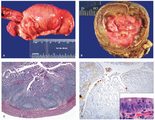
Figure 4-3. Lymphoid hyperplasia in small bowel and colon leading to intussusception of terminal ileum into the colon (A). The transverse section showing fleshy tissue in the invaginated segment representing lymphoid hyperplasia (B). Histology of the lymphoid hyperplasia in the colonic segment from the same case showing numerous lymphoid follicles with reactive germinal centers (C). The immunostain for adenovirus shows strong positivity in the overlying epithelium (D). Note the adenovirus nuclear inclusions (arrow) in the overlying epithelium (inset).


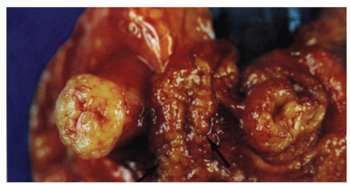
Figure 4-4. Focal lymphoid hyperplasia of the terminal ileum and appendix from a patient with symptoms of acute appendicitis. Gross appearances showing cobble stoning of ileal mucosa (arrow) and marked thickening of the appendix with luminal obliteration.


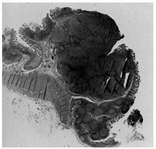
Figure 4-5. Focal lymphoid hyperplasia of the terminal ileum and appendix. Note the follicular hyperplasia of Peyer’s patch in the terminal ileum, on the right, extending into the appendix.


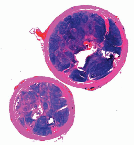
Figure 4-6. Focal lymphoid hyperplasia of the appendix. Lower-power photomicrograph of a cross-sectional cut demonstrating marked mucosal thickening, follicular hyperplasia, and luminal obliteration.


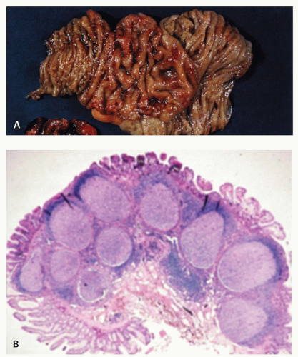
Figure 4-7. Focal lymphoid hyperplasia of the duodenum. A: The mucosal folds are focally thickened and edematous. Additionally, there are numerous nodules in both the edematous and nonedematous mucosa. B: Low-power photomicrograph of part (A) illustrating hyperplastic lymphoid follicles in the mucosa and submucosa. (Courtesy M. Ranchod, M.D.)


Localized Lymphoid Hyperplasia of the Rectum

Localized lymphoid hyperplasia of the large intestine, other than in the ileocecal area, appears to be located almost exclusively in the rectum.71, 72 This lesion is also known as benign lymphoid polyp or rectal (anal) tonsil.73 It may occur in all age groups but is most common in the second to fifth decades. The lesion has also been described in children.49, 74 Patients with benign lymphoid hyperplasia have a variety of symptoms, which include rectal bleeding, altered bowel habits, anal discomfort, and prolapse of a rectal mass. Many patients have associated anorectal lesions, such as hemorrhoids, anal fissures, and colonic carcinoma, which may contribute to their symptoms. Most patients have no evidence of nodular lymphoid hyperplasia elsewhere in the colon.

Pathology Localized lymphoid hyperplasia of the rectum usually affects its lowermost portion.49 About 60% to 80% of the lesions present as single polyp, and when multiple they are usually fewer than six. However, the polyps may be numerous on occasion and may impart a cobblestone appearance to the rectal mucosa.75 They are most commonly sessile, have a smooth surface, and have a pale yellow or white surface on endoscopy. They range from a few millimeters to 5 cm in diameter.49, 71, 73

A heavy lymphoid infiltrate is present in the lamina propria and submucosa. Large follicles with prominent germinal centers are always present, although they may be difficult to identify in poorly prepared tissue (Figs. 4-8 and 4-9). Eosinophils and plasma cells are not prominent. The majority of polyps are covered by intact, although at times attenuated, colonic epithelium.49, 71, 73 Low-lying polyps may be covered by anal squamous epithelium. The lymphoid nodules straddle the mucosa and submucosa and are therefore in a location similar to the solitary lymphoid nodules found in the normal colon. The muscularis mucosae show irregular proliferation and may be focally
absent. Focal lymphoid hyperplasia of the rectum is readily differentiated from malignant lymphoma (see the discussion of lymphoid hyperplasia of terminal ileum). It should also be kept in mind that malignant lymphoma is exceedingly rare at this site.13 As discussed in the preceding section, an immunodeficiency syndrome should be excluded.


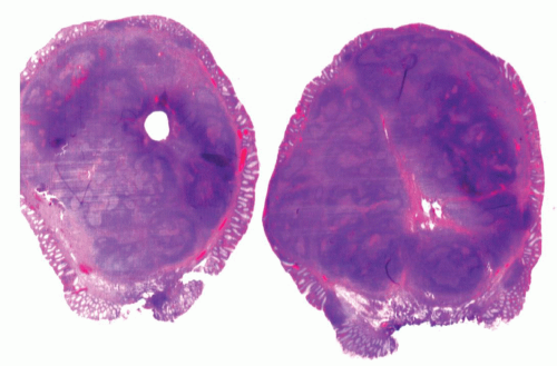
Figure 4-8. Focal lymphoid hyperplasia of the rectum (lymphoid polyp) demonstrating the polypoid appearance of the lesion. The lesion is covered by intact, focally attenuated colonic epithelium and contains large lymphoid follicles with prominent germinal centers.

Lymphoid polyps of the rectum should be well sampled for diagnostic purposes. If the lesions are multiple and the patient is asymptomatic, complete excision is not necessary, since the lesions may regress spontaneously. The incidence of local recurrence is low even after incomplete excision.49, 73


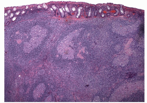
Figure 4-9. Microscopic appearance of rectal lymphoid hyperplasia showing numerous lymphoid follicles with germinal centers. Note that the infiltrate tends to push the crypts apart.


Diffuse Nodular Lymphoid Hyperplasia of the Intestine

Diffuse lymphoid hyperplasia in the small intestine can be either associated with or without hypogammaglobulinemia.

Diffuse nodular lymphoid hyperplasia with hypogammaglobulinemia. This disorder occurs primarily in patients with underlying congenital or acquired
immunodeficiency syndromes (see also Chapter 5).

Commonly associated immune disorders are CVID (late-onset hypogammaglobulinemia), selective IgA deficiency, and X-linked immunoproliferative disorder.76, 77, 78


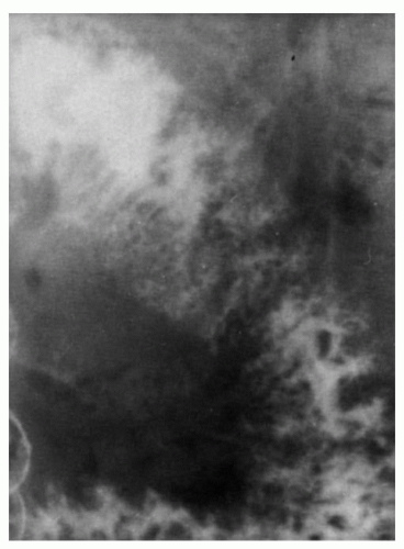
Figure 4-10. Nodular lymphoid hyperplasia with hypogammaglobulinemia: x-ray appearances. The barium radiograph is characterized by numerous nodular translucencies representing the mucosal lymphoid nodules.


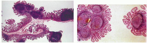
Figure 4-11. Low-power view showing hyperplastic mucosal lymphoid follicles. The mucosal nodules are composed of either (A) single or (B) clusters of hyperplastic lymphoid nodules.

Pathology The mucosa of the small intestine is studded with closely packed sessile or polypoid nodules measuring <5 mm in diameter. On x-ray, this produces a very striking picture characterized by numerous filling defects (Fig. 4-10). Rarely gastric and colonic involvement may occur.49, 79

The lesions consist of one or a cluster of hyperplastic lymphoid nodules, which are confined to the lamina propria and superficial submucosa (Fig. 4-11). They often produce shortening and blunting of the overlying intestinal villi. A decrease or absence of plasma cells is often noted in the lamina propria throughout the GI tract in CVID. Patients with selective IgA deficiency show absence of IgA immunoreactive plasma cells in the lamina propria in the background of normal IgG- and IgM-positive plasma cells. Untreated patients frequently have a superimposed parasitic infection, especially giardiasis. In rare instances, lymphoma has supervened.80, 81 (For a more detailed discussion, see the description of “Immunoeficiency Disorders” in Chapter 3.)

Diffuse nodular lymphoid hyperplasia without hypogammaglobulinemia. Diffuse nodular lymphoid hyperplasia of the intestine can occur in the absence of hypogammaglobulinemia and may be more frequent than that associated with late-onset hypogammaglobulinemia.82, 83, 84, 85 In a study of 1,000 consecutive autopsies, Robinson et al.85 found 30 cases of nodular lymphoid hyperplasia. The small intestine alone was involved in 13% of the cases, the large intestine alone in 40%, and both in 47%. The patients died of a variety of causes; none had GI symptom, giardiasis, or hypogammaglobulimia.

Nodular lymphoid hyperplasia of the colon has been observed in children undergoing barium enemas for a variety of GI complaints. It is clearly a common incidental finding that is unrelated to the patient’s symptoms.82, 84, 86 Serum immunoglobulin levels and T- and B-cell function studies found to be normal.82, 84, 85

It is likely that the lymphoid nodules represent hyperplasia of the solitary lymphoid follicles normally present in large numbers in the GI tract resulting in the characteristic radiologic and gross findings.82, 86, 87 Radiologically the lesions show umbilication of the nodules, which is a helpful diagnostic clue.82 One or more of the crypts overlying the lymphoid follicle are widened and elongated, allowing them to trap barium and produce the umbilication seen radiologically.

Pathology The lesions are grossly visible as pale yellow or white mucosal nodules and normally measure up to 0.4 cm in diameter, although rarely they may be as large as 2 cm (Figs. 4-12 and 4-13). Histologically, the mucosal nodules are similar to those seen in nodular lymphoid hyperplasia with hypogammaglobulinemia except that the lamina propria contains normal or increased numbers of plasma cells.

Clinical Implications and Differential Diagnosis of Diffuse Nodular Lymphoid Hyperplasia The clinical importance of diffuse lymphoid nodular hyperplasia lies in the recognition of the underlying immune disorder, differentiation from lymphoma, and increased risk of developing GI neoplasms, primarily lymphoma and carcinoma.81, 88 It is also clear that diffuse nodular lymphoid hyperplasia may also occur in the small and large intestines in patients without associated hypogammaglobulinemia and giardiasis. In some cases, it may represent underlying food allergy.89, 90 It may be found incidentally during the radiologic examination of the GI tract or at autopsy and should not be immediately incriminated for any GI symptoms the patient may have.


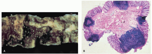
Figure 4-12. Nodular lymphoid hyperplasia of the large intestine. A: Showing mucosa studded with small, sessile nodules. B: Showing hyperplastic mucosal lymphoid follicles.

Nodular lymphoid hyperplasia of the ileum has also been described in patients with familial adenomatosis coli and Gardner’s syndrome, where it may give rise to multiple filling defects in the colon and occasionally in the ileum. This possibility should be considered with the usual small white bumps of the terminal ileum that represent normal lymphoid follicles and are routinely seen at endoscopy. Occasionally, this finding has resulted in colectomy with first-degree relatives of patients with familial adenomatosis coli as the polyps were presumed to be adenomatous without a biopsy.91 This also becomes important in youngsters as the surgeon may raise the question of ileal involvement and consider resecting affected small bowel.92, 93 Since patients with nodular lymphoid hyperplasia without hypogammaglobulinemia are asymptomatic and may undergo spontaneous regression, treatment of these lesions is unnecessary.82, 86 Most importantly, diffuse nodular lymphoid hyperplasia needs to be distinguished from various lymphomas that may present as multiple lymphomatous polyposis of the GI tract (see later).94 However, patients with diffuse nodular lymphoid hyperplasia without hypogammaglobulinemia only rarely develop lymphoma. Kahn and Novis reported a patient with multifocal “histiocytic” lymphoma of the GI tract.95 Matuchansky et al. reported a patient with nodular lymphoid hyperplasia who developed a jejunal lymphoma.96 Their case is of interest because they demonstrated immunohistochemically a transition from a hyperplastic lymphoid follicle to a neoplastic one. It is of interest that on many occasions when intestinal follicular or marginal zone lymphoma cases are examined, one can find hyperplastic lymphoid follicles immediately adjacent to the lymphoma and sometimes they even show same B-cell clones by molecular genetic analysis.


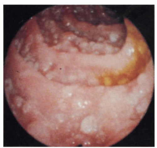
Figure 4-13. Nodular lymphoid hyperplasia of the large intestine: endoscopic appearance. The mucosa is studded with numerous pearly white nodules. (Courtesy S. Weiss, M.D.)


LYMPHOPROLIFERATIVE DISORDERS OF THE ESOPHAGUS

Primary lymphomas of the esophagus are extremely rare and account for <1% of all GI lymphomas.97, 98, 99, 100, 101 Many of these have been reported in the setting of immunodeficiency, particularly HIV infection.100, 102, 103, 104, 105, 106, 107 Secondary involvement of the esophagus from a mediastinal or gastric lymphoma is more frequently encountered in practice than a primary esophageal tumor. Clinically the patients present with symptoms that simulate carcinoma such as dysphagia, especially when the mass involves the distal esophagus.100, 108 The lesions tend to present as polypoid masses, prominent rugae, ulcers, or strictures.100, 104, 106, 109, 110 Multiplicity of lesions in this setting favors a lymphoma over a carcinoma. The majority of the lymphomas are of B-cell type. MALT lymphomas similar to those found elsewhere are also known to arise and tend to show similar histology (see the section on gastric MALT lymphoma).111 Histology reveals lymphoid follicles that are
surrounded by a diffuse infiltrate of CLC and a variable degree of plasma cell differentiation. Tumor cells infiltrating the overlying squamous epithelium are often present, suggesting epitheliotropism similar to lymphoepithelial lesions of gastric MALT lymphomas. Due to rarity of these lesions, it is unknown if the molecular genetics of these tumors is similar to MALT lymphomas at other sites. DLBCLs similar to their nodal counterparts are also known to arise in esophagus. Primary esophageal lymphomas including T-cell lymphoma, large-cell anaplastic Ki-1 lymphoma, Hodgkin’s lymphoma, and plasmacytoma have been reported in the literature and are exceedingly rare.97, 98, 106, 110, 112, 113


LYMPHOPROLIFERATIVE DISORDERS OF THE STOMACH

More than half of all primary and secondary GI lymphomas in the United States are of gastric origin. However, they represent only about 4% to 10% of all gastric tumors.114


MALT Lymphoma (Extra Nodal Marginal Zone Lymphoma)

Among the extranodal non-Hodgkin’s lymphomas, the stomach is the most commonly involved site in the Western countries, of which MALT-type lymphomas (MALTo-mas or MALT lymphomas) are the most frequent.1, 2

Pathogenesis. Understanding the pathogenesis of gastric lymphoma has been one of the major triumphs of this past century and continues to lead the way in our understanding of lymphomagenesis (Fig. 4-14). The association of H. pylori infection in patients with MALT lymphoma is currently well established.8, 115, 116, 117, 118, 119 Initial studies showed that H. pylori is associated with MALT lymphomas in 92% to 97% of the cases—a finding that is similar to gastric carcinoma—80% to 90% of which are also associated with H. pylori infection.119, 120 The prevalence of H. pylori infection in the general population in Western countries is about 30% but is much higher in developing countries where, in contrast to developed countries, children still tend to become infected early in life. Studies of archived gastric biopsies and sequential serological studies have shown that H. pylori infection precedes the development of gastric MALT lymphomas.33, 34, 119 With disease progression, the tumor growth tends to become independent of the organisms. At later stages, it is likely that the gastric environment becomes atrophic and no longer conducive to the survival of H. pylori, and either the organisms disappear or become too sparse to be visualized in biopsies. Thus, it is not surprising that the association of H. pylori with transformed MALT lymphomas (MALT lymphoma with a large B-cell component) is only 52% to 71%, while with purely DLBCLs (formerly high-grade MALToma) it is only 25% to 38%.121, 122, 123 Similarly, H. pylori can be demonstrated in 90% of cases when the tumor is limited to mucosa and submucosa, but in only 76% of the cases with submucosal spread of the tumor, and 48% of the cases when the tumor extends beyond submucosa.124


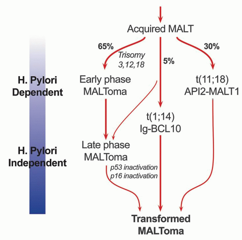
Figure 4-14. Schematic diagram showing the progression of H. pylori-driven B-cell proliferation leading to lymphoma.

The notion that the gastric mucosa (unlike small and large bowels) normally lacks organized lymphoid tissue is belied by experience in which small lymphoid aggregates can be found immediately above the muscularis mucosae even in children who have no indication of ever having had Helicobacter. However, the lamina propria shows only scattered lymphocytes and plasma cells. It has been hypothesized that H. pylori induces a lymphoid infiltrate associated with lymphoid follicles that is similar to the MALT of intestines and provides the background for the development of lymphoma.120 It has been shown in vitro that the tumor cells from a MALT lymphoma proliferate when incubated with heat-killed H. pylori, but die rapidly in the absence of H. pylori or when only chemical mitogens are used.125 This proliferative response also disappears when the T cells are removed from the culture.126 Addition of supernatant from another culture of unseparated tumor cells fails to restore cell proliferation, suggesting direct contact with T cells rather than secreted cytokines is important.126 This proliferative response to H. pylori not only appears to be strain specific for a given tumor, but varies among tumors from different patients. These observations suggest that the tumor is at least in part antigen dependent
and that H. pylori is the most likely antigen. It is also clear from these experiments that T cells play a vital role in the B-cell proliferation and that the B cells themselves are not H. pylori specific but make antibodies to a variety of autoantigens, including parietal cells and the proton pump that can result in gradual atrophy of specialized gastric mucosa. While the initial B-cell proliferation is H. pylori driven and polycloncal, subsequent molecular and genetic alterations lead to clonal expansion and autonomous growth. Whether H. pylori-specific factors play a role in the development of lymphoma is still somewhat controversial.127, 128, 129 The H. pylori strains, which accelerate the tumor growth, include the Cag A strain, a gene found in approximately 60% of H. pylori organisms, and the vac A (vacuolating cytotoxin) genotype.129, 130, 131 MALT lymphomas associated with H. heilmanni infection have also been reported.132 The cell of origin of MALT lymphomas is a B-cell that is identical to lymphocytes of the marginal zone of lymphoid follicles. These represent postfollicular B cells, and antigen selection occurs in some cases.133, 134, 135


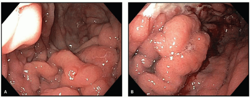
Figure 4-15. Lymphoma of the stomach: endoscopic appearances. A: Showing nodular mucosal lesions. B: Showing irregular mucosal masses, which may be indistinguishable from carcinoma. (Courtesy J. Nord, M.D.)

Clinical features. MALT lymphomas occur predominantly in the stomach in middle-aged or older patients although on rare occasions they have been reported in younger patients, as young as 7 years of age.136, 137 The tumor occurs more commonly in males (M:F = 1.5:1), the mean age being around 60 for males and 65 for females. The majority of the patients present with nonspecific symptoms that include epigastric pain, nausea, vomiting, or dyspepsia, similar to peptic ulcer disease. Patients with advanced disease or high-grade lymphomas tend to have signs and symptoms that mimic gastric carcinoma, such as early satiety, weight loss, hemorrhage, and anemia. Some cases may have a frank obstruction accompanied by a palpable mass.

Endoscopically, the tumors often have a varied and nondiagnostic appearance that includes severe thickened folds, superficial erosions, or deep ulcers (Fig. 4-15). In some instances the mucosa may show only minimal hyperemia or even appear normal. The lesions may be multifocal as shown by the microscopic involvement of apparently normally appearing mucosa in some cases. These findings are also reflected in the imaging studies, either CT scan or ultrasound-based evaluations. Endoscopic ultrasound (EUS) is a valuable imaging modality in evaluating the extent of involvement by lymphoma.138

Pathology. The gross appearance of the tumor is highly varied and includes flat, granular, or nodular mucosa; minimal to mild surface erosions; thickened folds; and ulcers or ill-defined diffusely infiltrative lesions (Fig. 4-16). However, the most common presentation resembles chronic active gastritis or gastric ulcers. Polypoid masses or ugly ulceroproliferative lesions are most commonly associated with advanced disease or high-grade tumors where the lesions could easily reach 8 cm in diameter. Typically, the lesions are localized to the mucosa and sub-mucosa, and the infiltrative growth results in either uniform or localized thickening of the wall. In some cases tumors are associated with the destruction of the full thickness of the gastric wall without an associated desmoplastic reaction and may lead to perforation. On cut section they have a characteristic white homogeneous fish-flesh appearance. As most of the lymphomas are low grade, detected early, and treated medically, fewer gastric resections with large lesions are encountered in current practice.

The most common site at presentation is the antrum. However, subsequently the tumor may spread throughout the stomach. Frequently it is multifocal,
although many foci may be microscopic in size. Once the infiltrate extends into the submucosa, it tends to separate the muscle fibers of the muscularis mucosae and either grows in a band-like manner, with a pushing border, or invades with an irregularly infiltrating margin. Eventually, the muscle undergoes atrophy, resulting in the marked weakening of the viscus and a propensity to perforation. The regional lymph node involvement is distinctly uncommon with superficial lesions and is seen once the tumor spreads to the muscularis propria or undergoes transformation to high grade.


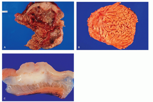
Figure 4-16. A: Gross photograph of a resection specimen of stomach showing large ulcerating lesion with marked thickening of the stomach wall. The cut section shows fleshy grey-white tumor diffusely involving the wall without any well-defined borders. B: Another specimen of stomach showing smaller ulcerating lesion with thickening of the mucosal folds around it. C: The cut section shows fleshy grey-white tumor infiltrating into the submucosa and muscularis propria without any well-defined borders.

The microscopic findings in MALT lymphomas are also varied and range from chronic active gastritis-like to typical nodal marginal zone lymphomas (Fig. 4-17). The majority of MALT lymphomas are limited to the mucosa and superficial submucosa, resulting in thickening of the mucosa with separation or gradual replacement of the glandular component. Initially, the crypts or glands are pushed apart from one another and upward from the underlying muscularis mucosae by the lymphoid infiltrate. Morphologically, the MALT lymphomas characteristically have histologic features that mimic normal MALT and are most commonly associated with a background of chronic active gastritis, but in which the monomorphous lymphoma appears to push aside the Helicobacter gastritis. The tumor cells tend to form monomorphous sheets of lymphocytes and infiltrate into the gastric pits, glands, or surface epithelium. The latter create the lymphoepithelial lesions, which are characterized by clusters of at least three or more atypical B cells infiltrating and destroying the foveolar or glandular epithelium (see Figs. 4-17 and 4-19). As normal IELs are usually T cells and therefore CD3 immunoreactive, the presence of these cells as B cells also aids with the diagnosis. However, if T-cell stains are carried out, MALT lymphomas appear to be T-cell driven so that the number of T cells is invariably as frequent as B cells to the point that a dearth of T cells should cast doubt on the diagnosis. The damaged epithelial cells often assume an eosinophilic/oncocytic appearance and may not be readily appreciable on H&E sections. In such situations, keratin stains are very useful to highlight the lymphoepithelial lesions. Occasionally, benign-appearing clusters of lymphocytes are found in the surface and crypt epithelium that represent the normal transmigrating lymphocytes. They differ from the lymphoepithelial lesions in that phenotypically they are T cells and they do not destroy the epithelium.139, 140

Lymphoid follicles are almost invariably present in MALT lymphomas and are frequently partially or totally surrounded by a sheath of atypical lymphocytes and plasma cells, which may be reactive or monotypic, occupying the mantle or marginal zones. These cells
tend to spread out from the marginal zone of the follicle to infiltrate the interfollicular zone and destroy the glands. Occasionally, these cells may also infiltrate the center of the follicle (follicular colonization), producing a vague nodular architecture (Fig. 4-18).141 This can be easily demonstrated by dendritic cell markers (CD21, CD23, or CD35) that highlight the background of dendritic reticulum cells in the colonized follicle. Another invariable finding in over 90% of these patients is the presence of H. pylori infection.


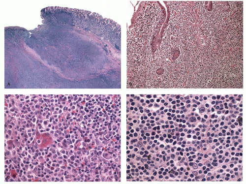
Figure 4-17. Microscopic appearance of gastric malt lymphoma. A: Low power showing extensive lymphocytic infiltrate with ulceration of mucosa and extension into the wall of the stomach. B: Lymphoepithelial lesions showing clusters of monocytoid B-cell infiltrating the glands. C: High power showing mixture of cells with many small lymphocyte-like cells, centrocyte-like cells, and plasma cells. D: High power showing large centroblasts/immunoblast-like cells interspersed among small lymphocyte-like cells.

Cytologically, the MALT lymphomas frequently consist of an admixture of various lymphocytic cell types, although one cell type may predominate in a given case. The commonly found types of lymphocytes are the following:

1. Small lymphocytes resembling normal mature lymphocytes but differing slightly in that they have somewhat enlarged irregular or wrinkled nuclei with less dense chromatin. MALT lymphomas with predominantly these types of cells may be difficult to distinguish from disseminated nodal small lymphocytic lymphoma/chronic lymphocytic leukemia (SLL/CLL) or occasionally a mantle cell lymphoma.

2. Lymphoplasmacytoid cells often containing intra-cytoplasmic immunoglobulin, commonly of the IgM class, with light chain restriction and Dutcher bodies (intranuclear periodic acid Schiff [PAS] positive inclusions). The presence of lymphoplasmacytoid cells in MALT lymphomas needs to be differentiated from the rare pure “small lymphocytic or plasmacytoid lymphomas” (immunocytomas of the Kiel classification), which are found in lymph nodes, spleen, bone marrow, and less frequently extranodal sites. Some of these cases may be associated with Waldenström’s macroglobulinemia. Some authorities consider this tumor to be distinct from MALT lymphomas.


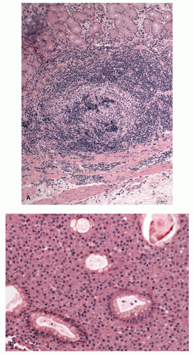
Figure 4-18. A: Microscopic appearance of follicular invasion by the marginal bone cells in a case of malt lymphoma of the stomach. B: Malt lymphoma of the stomach showing a predominant population of plasma cells. Often the plasma cells are localized in the superficial lamina propria.

3. CCLs are lymphocytes intermediate between small lymphocytic and small cleaved cell lymphomas, which have dense nuclei that are irregular in shape but are not as cleaved as the classical cleaved cell lymphoma. Variants of these cells, which have more abundant clear cytoplasm and well-defined cell margins, resembling monocytoid B cells, are frequently found.

In addition, large noncleaved lymphoblasts or immunoblasts may be scattered in between the small lymphocytes and do not imply high-grade transformation (Fig. 4-17C). Frequently there are large numbers of interspersed reactive T cells. Mature plasma cells are also frequently present and are most commonly distributed below the surface epithelium. The plasma cell component can be very prominent in some cases and may mimic a primary plasma cell disorder (see Fig. 4-18B). The cytoplasmic immunoglobulin in the lymphoplasmacytic cells has a perinuclear distribution and may be so abundant that it produces a “signet-ring”-like appearance mimicking a carcinoma.142 Since the immunoglobulin is preserved in formalin-fixed tissues and is PAS positive, it may cause further confusion with signet-ring cell carcinoma to the unwary, especially in small biopsies. On rare occasions metaplastic goblet cells from the surface or glandular epithelium may become pinched off by the lymphoid infiltrate and appear to lie isolated in the stroma, simulating signet-ring cell carcinoma.

Immunophenotype and molecular genetics. Immunohistochemically, the lymphomatous cells express surface and to a lesser extent cytoplasmic immunoglobulin, primarily IgM, which shows light chain restriction and rarely IgG or IgA. In addition, they are characteristically CD5, CD10, BCL1, and CD23 negative, but positive for the other B-cell markers (CD20, CD21, CD22, CD35, BCL2, CD79a, and CD23) and often CD43.8 In small biopsies, it may not be possible to perform all the markers, and one has to wisely choose a small panel that helps to differentiate MALT lymphoma from its mimics in a given case (see Table 4-5).

Many genetic and epigenetic abnormalities have been described in MALT lymphomas. However, most MALT lymphomas (about 70%-85%) do not have a specific translocation, yet there are four translocations that are specifically associated with MALT lymphomas and at the molecular level share a physiological role for BCL10 and MALT1 in antigen receptor-mediated NFkB activation.143 These include chromosomal translocations: t(11;18)(q21;q21), important because it predicts lack of response to Helicobacter eradication therapy, and t(1;14)(p22;q32) and t(14;18)(q32;q21) that seem to be specific for MALT lymphomas and have diagnostic and prognostic importance.144, 145, 146 Another more recently recognized translocation with MALT lymphomas is t(3:14)(p14.1q32) and suggests a role of FOXP-1. The proposed molecular mechanisms and pathogenesis of H. pylori-associated MALT lymphomas are shown in Figure 4-14. Other abnormalities include trisomies 3,12,18; p53 mutation/LOH; p15 and p16 promoter methylation; and fas mutation.147, 148, 149, 150, 151, 152, 153, 154

Translocation t(11;18)(q21;q21) In most of the translocation positive cases, t(11;18)(q21;q21) is the sole chromosomal abnormality.144, 145 This translocation causes reciprocal fusion of the API2 and MALT1
genes.155, 156, 157 The API2 is an apoptosis inhibitor, whereas MALT1 is involved in antigen receptor-mediated NFkB activation. The fusion transcript is consistently expressed in these cases and is believed to be oncogenic. Unlike wild-type API2 and MALT1, API2-MALT1 fusion product is a potent activator of NFkB. NFkB activation in turn leads to activation of other cytokine and growth factor genes that are important for cellular activation, proliferation, and survival, thus contributing to tumor development.158, 159, 160 This translocation is seen in about 15% to 30% of gastric MALT lymphomas.161 The same translocation has also been described in MALT lymphomas of other sites including skin and lungs. However, it is absent in nodal and splenic marginal zone lymphomas.162, 163, 164, 165 Cases of H. pylori gastritis also do not show this translocation. MALT lymphomas with this translocation do not respond to H. pylori eradication and more often spread to distant sites including regional lymph nodes.166 An initial report suggested that despite the association of t(11;18)(q21;q21) with adverse clinical features, the translocation is uncommonly found in cases with DLBCLs or so-called transformed MALT lymphomas, that is, low-grade MALT lymphomas with a large-cell component.167, 168 A subsequent study however shows that this translocation is seen with almost equal frequency in transformed MALT lymphomas or DLBCLs of the stomach.169 Even if found, H. pylori eradication treatment is undertaken together with other lymphoma therapy.


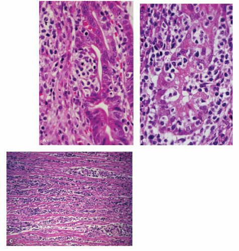
Figure 4-19. MALT lymphoma of the stomach. A,B: Medium-power photomicrographs showing infiltration of glands by the atypical lymphocytes. C: Lymphoma showing separation of the muscle fibers of the muscularis propria by the lymphomatous infiltrate.

Translocation t(1;14)(p22;q32) The second common MALT lymphoma associated translocation is t(1;14) (p22;q32). Cases positive for this translocation are often
associated with trisomies 3, 12, and 18, unlike t(11;18) (q21;q21).146 The translocation brings the entire BCL10 gene under the regulatory control of the Ig gene and deregulates its expression.143, 170 Earlier studies showed that although BCL10 activates NFkB, it is weakly proapoptotic.171, 172 However, subsequent studies have shown that BCL10 does not have proapoptotic activity in vivo.173, 174 It is essential for the development of B- and T-cell functions and specifically links the antigen receptor signaling to NFkB pathway.175, 176, 177 In response to the surface antigen receptor stimulation, BCL10 oligomerizes and binds to MALT1, leading to MALT1 oligomerizartion that in turn activates NFkB. In line with the physiological role of BCL10 in normal B cells, the protein is expressed predominantly in the cytoplasm of germinal center B cells.178 BCL10 is strongly expressed in the nuclei of MALT lymphomas with t(1;14)(p22;q32) or its variants.178 However, moderate nuclear BCL10 expression is also seen in about 50% of cases negative for this translocation and in almost all t(11;18)(q21;q21)-positive cases.161, 165, 179 Studies using BCL10 immunostains and interphase FISH analysis show that t(1;14)(p22;q32) is specifically associated with MALT lymphomas and not with other lymphoma subtypes.165 This translocation occurs in about 5% of gastric MALT lymphomas and has also been associated with MALT lymphomas of lungs.165 Gastric MALT lymphomas with this translocation are typically at advanced stages and unlikely to respond to H. pylori eradication.

Translocation t(14;18)(p32;q21) The third MALT lymphoma associated translocation t(14;18)(p32;q21) is different from the similar translocation seen in follicular lymphomas. In follicular lymphomas this translocation brings BCL2 at 18q21 in proximity to Ig heavy chain (IgH) locus. The same breakpoint in MALT lymphomas involves MALT1 instead of BCL2.180 It is proposed that IgH-MALT1 fusion results in activation of MALT1 that somehow stabilizes BCL10 in the cytoplasm. BCL10 in turn then leads to aberrant activation of NFkB pathway. This translocation has so far been shown to be associated with MALT lymphomas of the lungs and ocular adenxae, but not stomach.164, 180, 181

Translocation t(3;14)(p14;1q32) This is the more recently recognized MALT lymphoma-associated translocation that brings FOXP1 gene in proximity to Ig heavy chain gene and results in its deregulation.182 FOXP1 belongs to the Forkhead box family of winged-helix transcription factors that have diverse function in different cell types and plays an important role in B-cell development. This translocation was detected in a single case (1.7%, n = 59) of MALT lymphoma studied and seems to correlate with poor outcome and transformation to DLBCL.182, 183 This translocation has also been reported in isolated cases of gastric DLBCL.

Treatment and prognosis. These are typically slow-growing lesions that remain localized to the stomach for long periods (stage 1E), often more than 7 to 10 years.138, 184, 185 The majority of gastric MALT lymphomas at the time of presentation are confined to the mucosa and submucosa. There is a low incidence of mesenteric node involvement and extraintestinal dissemination, although in one study bone marrow involvement was found in 10% of the patients.5 Recurrence of these low-grade tumors occurs primarily in patients with an increased number of transformed lymphocytes (5%-10%) and involves the original site or other parts of the GI tract, mesenteric lymph nodes, or Waldeyer ring, rather than widespread extraintestinal dissemination.

The overall prognosis of these patients is excellent. As has been discussed earlier, remission of the majority of MALT lymphomas (67%-90%) occurs following the treatment of H. pylori.186, 187, 188 This is largely applicable to tumors that do not have a high-grade component and are localized to mucosa or superficial submucosa. Advanced tumors or those with a high-grade component have also been reported to respond to H. pylori eradication, but infrequently. In several large studies, 90% of patients survived for 5 years and 65% to 75% of patients for 10 years. Whether the patients are truly cured still remains to be determined. Relapses have been shown to occur in about 10% of patients.189 In some patients, the relapses have been associated with reinfection with H. pylori and successful remission following eradication of the infection again.

It is increasingly becoming clear that molecular genetics has an important role in predicting the behavior of gastric MALT lymphomas. Tumors with t(11;18) (q21;q21) translocation generally do not respond to H. pylori eradication, even though they are rarely associated with large-cell transformation. Chemotherapy (sometimes with radiation) and occasionally surgery are the options for cases that do not respond to H. pylori eradication. Surgical resection on its own has been associated with prolonged survival in some studies.190 Involvement of the resection margin and advanced stage are poor prognostic factors, but not when adjuvant chemotherapy is also used.191 These tumors invariably express CD20, and resistant cases or those with large-cell component have been successfully treated with rituximab; however, long-term outcome of such cases is still unknown.192 Irrespective of the treatment modality, the only independent prognostic variables are stage and tumor grade.191, 193, 194, 195, 196

Reporting MALT lymphomas. There is considerable variation in how pathologists report gastric MALT
lymphomas in biopsies. When the biopsies are adequate and the morphology and immunohistochemistry are typical, many are comfortable making the diagnosis, while some always hedge by stating “consistent with”. One of the problems with MALT lymphomas is the lack of a distinct immunohistochemical profile, and stains are largely used to exclude other lymphoma types (mantle, follicular, CLL etc). Presence of one of the MALT associated translocation should certainly help one in making a definitive diagnosis however, these are present only in a small proportion of cases and are still not readily available in all labs. Nevertheless, pathologists should still be comfortable making this diagnosis in the presence of appropriate histology with the knowledge that the first line of therapy is Helicobacter eradication followed by re-endoscopy and biopsy a few months later. If the morphology and immunohistochemistry are all correct, but Helicobacter cannot be demonstrated or the biopsies are too small, reporting them as “atypical lymphoid infiltrate” and suggest follow-up biopsies for a full lymphoma work-up with flow-cytometry, molecular assays and serology for Helicobacter.

Follow-up of patients with treated MALT lymphoma. Follow-up of these patients after therapy involves periodic endoscopic and EUS examinations and biopsies.197 It should be remembered that where there has been an endoscopically visible lesion associated with Helicobacter, following its eradication the primary purpose of the endoscopy is to ensure that the lymphoma is regressing or at least not increasing in size. The latter would suggest incomplete eradication of Helicobacter, a translocation associated with lack of response to Helicobacter therapy or a large-cell component. Biopsies would therefore be expected to contain residual MALT lymphoma, and the demonstration that this is the case is unnecessary.

Role of the pathologist. The role of the pathologist is therefore not to just confirm the presence of residual lymphoma, which is a given as these cells are long-lived and may persist for long periods of time following the withdrawal of the antigenic stimulus, but to also

1. Identify the presence of a large-cell component when present.

2. Identify Helicobacter if present (coccoid forms are not acceptable and are much more likely to be oral or enteric in origin). It should also be recalled that patients on long-term PPIs may allow oral organisms to grow in their stomachs. These are usually polymorphous and can include fungi. Helicobacter therefore have to be photographable (stand up in court) but are important to identify as the lymphoma will not regress until eradication is complete.

3. A useful stain is Mib-1 to evaluate the proportion of cells in non-G0 phase of cell cycle, especially if this can be compared with a pretreatment biopsy, as the proliferation usually falls off markedly once the antigenic stimulus is withdrawn. In the absence of a large-cell component, a persistently high Mib-1 rate suggests persistent Helicobacter, but if these are really absent, one needs to carry out translocation studies. Although monoclonal antibodies to one of the translocation products and also BCL10, which has been suggested as a surrogate marker, are helpful, at the time of writing these are not reliable enough to be part of routine practice. However, in some centers it is thought that there is an economic advantage in looking for the translocations, especially the t(11;14), as it can be predicted that these will not respond to Helicobacter eradication, so the patients will need chemotherapy (which does not preclude Helicobacter eradication).

The long-term follow-up biopsies from patients continuing to respond to Helicobacter eradication therapy ultimately show that the number of biopsies with MALT tend to diminish and disappear over time. At this stage biopsies often show an empty-looking lamina propria with rare B cells or plasma cells.198 Sometimes small aggregates of normal-appearing small lymphocytic B cells and scattered T cells are also evident and do not imply residual/recurrent lymphoma. Studies using PCR and Ig gene rearrangement have shown that residual clones can be identified in many cases without any histologic evidence of residual disease.197, 199 It has been shown that histologic remission may take many months or years (median 33, range 0-65 months), while molecular remission takes longer.198, 200, 201, 202 At present, the significance of the persistent molecular abnormality in the setting of clinical and histologic remission remains unclear. However, current data also do not justify further treatment of persistent monoclonality by molecular assays in the absence of histologic or clinical disease.

Lastly, it should be noted that the patients with MALT lymphomas also seem to have an increased incidence of gastric adenocarcinoma.203 This often arises in the distal stomach and appears to occur irrespective of the type of therapy for the lymphoma.

Differential diagnosis and practical approach to the difficult diagnosis. Whenever there is dense lymphocytic infiltrate in the gastric mucosal biopsies, the differential diagnosis includes a lymphoma. The problems often one faces in this situation are as follows:

1. When the infiltrate is polymorphous, the issue is to differentiate between H. pylori-associated chronic active gastritis and MALT lymphoma.


2. When only a small focus of B lymphocytes invading a crypt forming an isolated lymphoepithelial lesion is identified (mini-MALT lymphoma).

3. When the lymphocytic infiltrate is clearly neoplastic, the issue is the correct subclassification of the lymphoma.

Chronic Gastritis versus Malt Lymphoma This situation is frequently encountered in routine practice. The lymphocytic infiltrate in lymphoma tends to be more destructive and expansible, with extensive permeation between the glands. The presence of significant numbers of small cleaved cells (CLC or monocytoid B cells) also supports the diagnosis; however, in some cases these features are not readily evident. The presence of convincing lymphoepithelial lesions strongly favors the diagnosis of MALT lymphoma, although one needs to be careful as they can also be seen in florid H. pylori gastritis (usually single, consist of <3 cells and rarely destructive) as well as other types of lymphomas. The presence of Dutcher bodies or intranuclear inclusions in more than a few plasma cells is also suggestive of lymphoma; however, similar to other sites mere presence of these inclusions by itself is not diagnostic. In reactive lymphoid infiltrate the B cells are typically organized in nodules even though the lymphoid follicles may not be easily evident, and the T cells reside in the interfollicular zones. This pattern can be easily appreciated with CD20 and CD3 immunostains. If CD20 stain shows a diffuse densely packed infiltrate of B cells with limited number of interspersed T cells, this pattern supports a diagnosis of lymphoma. Light chain restriction using immunostains for κ and λ light chains may be helpful in demonstrating a monotypic Ig supporting the diagnosis of lymphoma. However, interpretation of these immunostains performed on paraffin sections is not always straightforward unless overtly plasmacytic. In addition, a polyclonal infiltrate of chronic gastritis often coexists in the background, and one needs to carefully interpret the stain in the atypical cells. Coexpression of CD43 by CD20-positive B cells also supports the diagnosis of lymphoma, although a normal immunophenotype does not exclude this possibility. Detection of a clonally rearranged Ig gene by PCR in the presence of atypical infiltrate is diagnostic of lymphoma. However, a diagnosis of lymphoma should never be based solely based on this finding. Demonstration of any of the MALT lymphoma-associated translocations as discussed above using RT-PCR or FISH is also diagnostic of lymphoma. This not only provides an evidence of a clonal B-cell expansion but represents a MALT-specific abnormality that also has a prognostic significance. If the studies are inconclusive or equivocal, and the biopsy shows an evidence of H. pylori gastritis, repeat biopsies can be suggested following the treatment for H. pylori. In such a setting, it would be appropriate to use the designation “atypical lymphocytic infiltrate of uncertain nature or significance.” If in a similar setting H. pylori is negative, repeat biopsy with more sampling should be suggested sooner than later. If biopsies are performed for confirming a diagnosis of lymphoma, it is advantageous to have additional samples for flow cytometry and molecular studies obtained at the outset.

Mini-MALT Lymphomas In Helicobacter-positive patients, one sometimes comes across a focus of monotonous lymphoid cells surrounding one or two pits that contrasts with the plasma cell rich polymorphous infiltrate of the background gastritis. Further, there may be a focus of IELs mimicking a lymphoepithelial lesion, although these are rarely destructive. In our experience, some of them even show B-cell clonality based on immunoglobulin gene rearrangement studies. These likely represent the earliest stages of MALT lymphoma and likely disappear following H. pylori eradication. Since prospective studies looking at the natural history of such lesions are lacking, a cautious approach is warranted. We do not report these as MALT lymphomas but do not ignore them completely either, stating that a lymphoepithelial-like lesion is present associated with the Helicobacter gastritis and that this will likely disappear following Helicobacter eradication therapy. While calling such lesions as outright MALT lymphoma in the absence of any supportive molecular evidence may be an overkill, a suggestion in the report that Helicobacter eradication therapy should be considered to ensure these lesions do not progress is justified. Given the frequency of these lesions if carefully looked for and the rarity of MALT lymphomas, it is likely that progression of these lesions to overt MALT lymphoma is extremely rare.

Subtyping of a Small Lymphocytic Lymphoma In some cases, the lymphomatous nature of the infiltrate is obvious on histology due to the monotonous appearance of the infiltrate, its destructive nature, or cellular anaplasia. However, not all lymphomas of the stomach are MALT lymphomas, and proper subtyping of the lymphoma is essential as the clinical outcome of similar-appearing lesions is vastly different. The difficulties often arise due to small sample size or crush artifacts that obscure the histologic details and pattern. The entities that often come into the differential diagnosis include SLL/CLL, follicular lymphoma, and mantle cell lymphoma. The presence of lymphoepithelial lesions, though helpful, is not diagnostic in this situation as they have been described with a variety of other primary or secondary lymphomas of the stomach. Immunophenotyping and demonstration of specific translocations are
extremely valuable in this setting. However, if the sample is small and the findings are inconclusive, additional biopsies for diagnostic workup should be requested. CLL/SLLs have often involvement of peripheral blood, and knowledge of the peripheral blood findings could be helpful.

One of the most difficult and controversial issues is the recognition of DLBCL or, as formerly called, “large-cell/high-grade transformation” of MALT lymphomas (also see later). As defined in the WHO classification, the presence of sheets of large B cells is required to make this diagnosis; however, what constitutes a “sheet” is unclear.29 Scattered centroblasts and immunoblasts intermingled with the infiltrate are not uncommon in MALT lymphomas, and it has been suggested that more than 10% of large cells should prompt a diagnosis of DLBCL. More than 30% staining with Ki67 has also been suggested to support the diagnosis of DLBCL.


Diffuse Large B-cell Lymphoma of the Stomach

This group comprises MALT lymphomas with high-grade transformation and primary de novo large B-cell lymphomas of the stomach.8, 204 It had been lumped under high-grade MALT lymphomas in the past. The recommendation of the WHO classification is not to use the term high-grade MALT lymphoma,28 although if one of the recognized MALT translocations or other appropriate molecular abnormalities is present or the patient had a previous low-grade gastric MALT lymphoma, it is difficult to come to any other conclusion. DLBCL can be divided into the following categories:

1. Low-grade MALT lymphoma with a minor DLBCL component comprised of only occasional clusters of large cells.


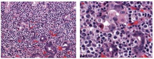
Figure 4-20. A: Appearance of DLBCL of the stomach. The morphology resembles that of an extranodal DLBCL of the stomach. B: There are numerous large cells in a background of H. pylori-positive MALT-type lymphomas and lymphoepithelial lesions are still clearly visible.

2. Equal admixture of DLBCL and MALT lymphoma components.

3. DLBCL with a small component of MALT lymphoma.

4. DLBCL without a MALT lymphoma component.

The relative proportion of each group varies in different studies largely due to lack of uniform diagnostic criteria and variation in classification.205, 206, 207 The presence of identical Ig heavy chain rearrangement in both components strongly suggests transformation of the low-grade MALT lymphoma to the large-cell component in many cases.208, 209 Occasionally, genetic difference between the two components suggests an independent evolution or dedifferentiation. Cases of purely DLBCL may represent cases of MALT lymphomas, where the large-cell component may have completely overgrown the low-grade tumor or a de novo large B-cell lymphoma (Fig. 4-20).194 Nevertheless, an increasing large-cell component is associated with a reduced positivity for H. pylori, increasing non-responsiveness to H. pylori eradication, and higher stage, and hence the rationale to lump them together as DLBCL seems justified.

Only gold members can continue reading. Log In or Register to continue

Stay updated, free articles. Join our Telegram channel

Jul 20, 2016 | Posted by in GASTROENTEROLOGY | Comments Off on Lymphoproliferative Disorders of the Gastrointestinal Tract

Full access? Get Clinical Tree

Get Clinical Tree app for offline access