, Ivan Damjanov1 and Ryan M. Taylor2
(1)
University of Kansas School of Medicine, University of Kansas, Kansas City, KS, USA
(2)
Division of Gastroenterology and Hepatology, University of Kansas School of Medicine, Kansas City, KS, USA
The normal liver weighs 1.2–1.6 kg and is located in the right upper abdominal quadrant. Traditionally it has been divided into a left and a right lobe, separated one from another by the falciform ligament. A functional division into a right and a left hemiliver, each of which is subdivided into four smaller segments is based on the blood supply. Subdivision of the liver into eight segments, each of which has its own blood supply and segmental bile ducts, is more useful for radiologic imaging purposes and surgical interventions.
Each liver segment is made of lobules, formed of liver plates and sinusoids radiating from a central vein, also known as the terminal hepatic venule (THV) (Figs. 1 and 2). At the periphery of the lobules the liver contains portal tracts, comprising branches of the portal vein, hepatic artery and bile ducts, surrounded by small amounts of fibrous connective tissue (Figs. 3 and 4). From the functional point of view, this anatomic concept of lobules has been replaced by the physiologic concept of acini, which represent the basic functional units of the liver. In the acini the liver cells are centered around the portal tracts, whereas the terminal hepatic venules are placed at their periphery. The acini are divided into three zones (Fig. 5):
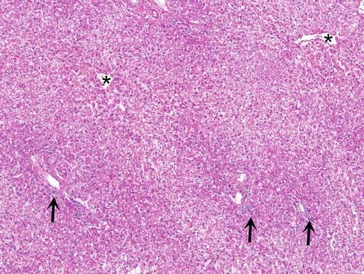
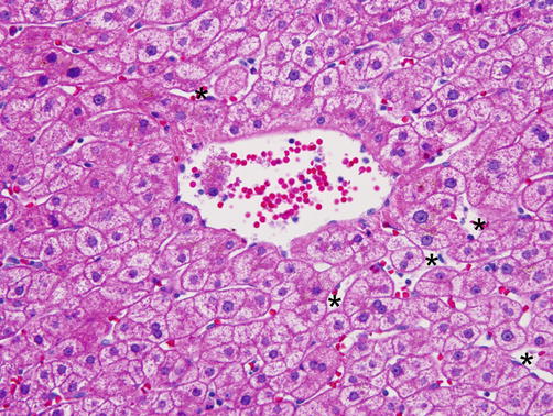
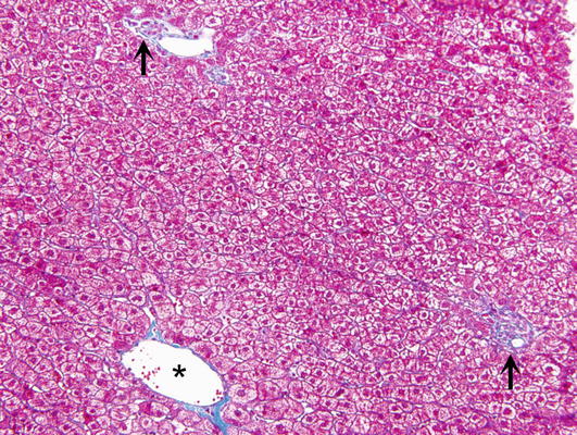
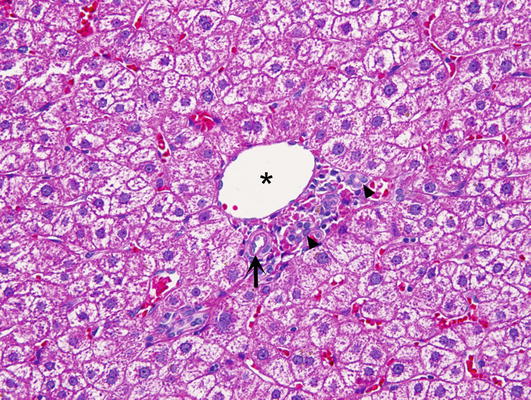
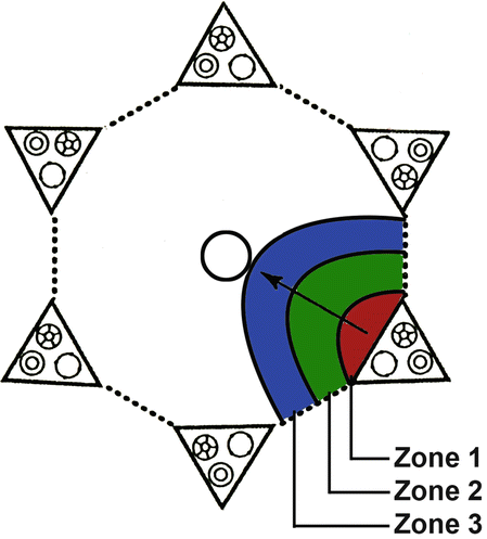
Fig. 1.
Normal liver. It is composed of lobules which are demarcated one from another. The main hallmarks are terminal hepatic venules (asterisks) and portal tracts (arrows)
Fig. 2.
Normal centrilobular zone 3. Normal liver cells are arranged around the terminal hepatic venule which contains some blood in its lumen. Liver cells are tightly attached one to another on their lateral side while facing the lumen of sinusoids on their vascular pole. In the sinusoids (asterisks) one may see the spindle shaped nuclei of endothelial and Kupffer cells, and occasional lymphocytes, which have round nuclei
Fig. 3.
Normal liver. Trichrome stain highlights the collagenous connective tissue in two portal tracts (arrows) and the wall of a terminal hepatic venule (asterisk)
Fig. 4.
Normal periportal zone 1. A small portal tract contains a branch of the portal vein (asterisk), a branch of the hepatic artery (arrow), and bile ducts (arrowheads)
Fig. 5.
Schematic presentation of two hepatic lobules and the concept of the acinus. The acinus is divided into three zones: periportal zone 1 (red), centrilobular zone 3 around the terminal hepatic venule (blue), and the zone 2 (green) between the zone 1 and 3. Portal tracts, each containing a bile duct, an artery, and a vein, are represented by triangles. The circles in the center of these two lobules represent the terminal hepatic venules. The arrows indicate the direction of the blood flow from the portal tracts toward the terminal hepatic venule
Zone 1—located closest to the portal tract. Best oxygenated and closest to the supply of nutrients and toxins from portal and arterial blood. Also known as periportal part of the lobule or acinus.Related posts:

Stay updated, free articles. Join our Telegram channel

Full access? Get Clinical Tree


