Lateral Decubitus Approach to Minimally Invasive Low Anterior Resection

Chapter 27 Lateral Decubitus Approach to Minimally Invasive Low Anterior Resection


imageThe videos associated with this chapter are listed in the Video Contents and can be found on the accompanying DVDs and on Expertconsult.com.


The questions and controversies that have surrounded the minimally invasive low anterior resection for rectal cancer have, to a large extent, been answered or refuted. These issues involve the learning curve, patient safety, cost, operative time, and whether it is possible to perform an oncologically sound resection with a laparoscopic approach. Recent data have suggested that the number of laparoscopic low anterior resections a surgeon needs to perform to obtain reasonable technical proficiency is in the range of 15 to 80. Studies on perioperative outcome have shown that laparoscopic low anterior resection has acceptable (perhaps improved) morbidity and mortality compared with the open procedure.


Pathologic specimen analysis and short-term follow-up so far have demonstrated that minimally invasive rectal resection is oncologically sound; long-term survival and recurrence data currently are accruing. Objections concerning the potential for port-site metastasis in laparoscopic colorectal resection have not been borne out. Laparoscopic colorectal resection has been associated with increased operative time and equipment costs compared with open resection. On the other hand, the former approach has been associated with an earlier return of bowel function, an earlier return to work, decreased pain scores and pain medication requirements, and a shorter length of stay.


The traditional approach to the laparoscopic low anterior resection has been with the patient supine, typically in a modified lithotomy position. The technical obstacles to this approach included difficulty in dissection of the transverse colon and the splenocolic ligament. On the other hand, the lateral approach in the decubitus position provides excellent visualization of the transverse and left colon and allows improved access to the splenic flexure. This permits easy mobilization of the colon without requiring excessive traction, while minimizing the number of ports needed. A low rectal dissection can be performed, and the colorectal anastomosis is constructed with a traditional end-to-end stapler technique.


Preoperative evaluation, testing, and preparation


The preoperative management of patients undergoing laparoscopic low anterior resection varies according to the surgical indication. Candidates must be medically fit to undergo general anesthesia, possibly prolonged, and must be able to withstand the physiologic challenges that pneumoperitoneum imposes. The following testing and preparation can be applied to any patient having this procedure, except that endoscopic ultrasound (EUS) and magnetic resonance imaging (MRI) usually are reserved for the patient with rectal cancer.


Patient positioning in the operating suite


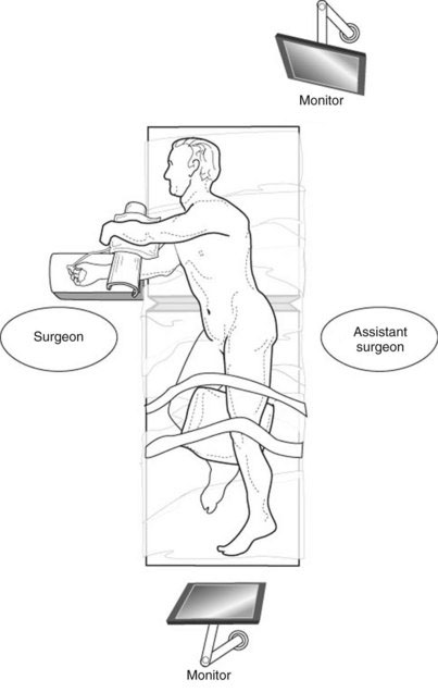
The patient is placed in the right lateral decubitus position (Fig. 27-1). The right leg is flexed 30 degrees at the hip, and the left leg is extended with a pillow placed between the knees. The buttocks are positioned toward the edge of the table. Sequential compression devices are placed on both legs, and the patient is taped securely to the table. The right arm is extended over an arm board, and the left arm is positioned over a pillow on a Mayo stand. An axillary roll is placed under the right axilla. The arms are properly cushioned and supported to avoid a perioperative neurapraxia. The table is flexed and the kidney rest may be raised, extending the left flank and maximizing the working space. The operative field is prepared and draped to allow exposure from the left nipple down to the pubic symphysis, and from the umbilicus to the posterior axillary line. The anus must be accessible under the drapes so as to place the EEA device, as if performing a colonoscopy. Two video monitors are required—one at the head of the table to the patient’s left, the other at the foot of the table.


Positioning and placement of trocars


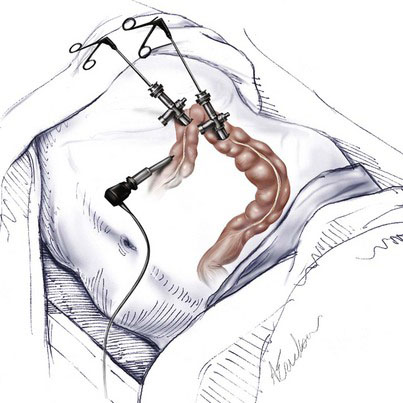
Laparoscopic low anterior and rectal resection in the right lateral decubitus position requires three trocars, with the option of a fourth (Fig. 27-2). A 10-mm trocar is inserted in the left midclavicular line, 2 to 3 cm above the level of the umbilicus. This serves as the optical port. A 5-mm trocar is inserted in the left subcostal region at the level of the anterior axillary line. This serves as the working port, through which the retraction and dissection instruments are placed. It can be converted to a 10-mm port if needed later in the operation. A 5-mm trocar is inserted under the left 11th rib at the level of the midaxillary line. This is used for the retraction and dissection instruments. If a fourth port is needed, it may be placed in the suprapubic region. A 10-mm 30-degree laparoscope is used for the procedure.


Operative technique


Mobilization of the colon and rectum is performed in two stages. The colon is mobilized in the first stage. This is best performed with the surgeon standing on the right side of the table and facing the upper monitor. The patient is placed in a 30-degree reverse-Trendelenburg position. This allows gravity to aid in the retraction of the colon, which facilitates the dissection of the splenic flexure and identification of the left ureter. Most of the planes are avascular, and dissection can be safely performed with an ultrasonic or bipolar dissector, or with electrocautery (hook or scissors).


The operation is begun by retracting the greater curvature of the stomach anteriorly to enter the lesser sac. The left hand retracts the mid-curvature, and the thin avascular layer of omentum that connects the stomach to the transverse colon (also known as the gastrocolic ligament) is entered bluntly or with an energy source. Care should be taken to ensure that the dissection does not extend into the mesocolon, which could cause bleeding or colonic ischemia. Depending on the transparency of the tissue, the division of the gastrocolic ligament proceeds toward the splenic flexure with electrocautery or with a dissecting-sealing device.


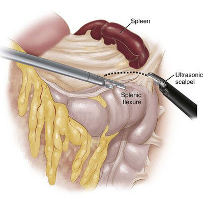
The advantage of the lateral decubitus approach becomes apparent during the dissection of the splenic flexure. As the colon is retracted inferiorly and medially with the right hand, the splenocolic ligament is transected (Fig. 27-3). This may best be performed using a sealing device because this ligament may contain small vessels. The flexure should fall away naturally from its attachments. The white line of Toldt then can be opened inferiorly toward the sigmoid colon. Careful attention to the planes of dissection must be maintained to avoid entering the lateral attachment of Gerota fascia, which would medialize the left kidney and reduce exposure of the operative field.


< div class='tao-gold-member'>

Stay updated, free articles. Join our Telegram channel

Jul 20, 2016 | Posted by in GASTOINESTINAL SURGERY | Comments Off on Lateral Decubitus Approach to Minimally Invasive Low Anterior Resection

Full access? Get Clinical Tree

Get Clinical Tree app for offline access