Laparoscopic Truncal Vagotomy with Antrectomy and Billroth II Reconstruction
Kfir Ben-David
George A. Sarosi Jr
Recent advancements in pharmaceutical therapy and endoscopic treatments have significantly decreased the incidence of gastric surgery for peptic ulcer disease (PUD). With the advent of proton pump inhibitors, histamine receptor blockers, and multidrug therapy treatment for Helicobacter pylori, previously common operations for PUD have become very infrequent. Consequently, medical progress and new surgical innovations have completely transformed our approach to patients with benign gastric and PUD in need of surgical treatment. With the use of flexible endoscopy and minimally invasive surgical approaches the diagnosis and surgical treatment of PUD has resulted in less invasive surgical treatments resulting in shorter hospital stay, less overall cost, and quicker return to base-line activity while being able to maintain the same surgical technique without patient compromise.
The laparoscopic approach to PUD with truncal vagotomy, antrectomy, and Billroth II reconstruction is theoretically possible in nearly all patients with benign surgical disease who have failed medical and endoscopic treatments. Many minimally invasive gastrointestinal surgeons feel that PUD should be preferentially treated via a laparoscopic approach since it is associated with less intraoperative blood loss, earlier return of bowel function, less usage of analgesics, and a shorter postoperative hospital stay. Despite these strong case series favoring a laparoscopic surgical approach, there is a relative paucity of randomized clinical trials addressing this issue, likely due to the decreasing incidence of PUD and the infrequent need for elective surgical treatment. However, a number of patient characteristics represent relative indications and contraindications to the laparoscopic approach.
Even though the natural course of PUD is changing and intractable disease and pyloric obstruction as an indication for surgery is decreasing, the incidence of bleeding and perforation remains constant. Treatment of H. pylori and cessation of nonsteroidal anti-inflammatory drugs is imperative prior to any surgical procedure for PUD. Although there have been a number of case reports describing laparoscopic surgical treatment with antrectomy, vagotomy, and Billroth II reconstruction for acutely perforated or bleeding PUD, many of these procedures were performed by very skilled laparoscopic surgeons. Furthermore, prior upper abdominal midline incisions and previous gastric and/or esophageal surgery are a relative contraindication to a laparoscopic truncal vagotomy, antrectomy, and Billroth II reconstruction. Mesh placement in the upper midline makes laparoscopic resection and reconstruction much more difficult. Liver cirrhosis and portal hypertension are also a contraindication to laparoscopic truncal vagotomy, antrectomy, and Billroth II reconstruction because of the risk of bleeding from gastric and esophageal varices. However, with increased experience utilizing laparoscopic approaches to gastric resection and reconstruction, patients with advanced age and its associated chronic medical conditions are not generally contraindications.
Most of the preoperative evaluation is directed toward ensuring that the patient is adequately prepared for anesthesia. As a result, a careful assessment of the patient’s fitness to undergo general anesthesia represents a major portion of the preoperative assessment. Because truncal vagotomy, antrectomy, and Billroth II reconstruction will almost always be performed under elective circumstances, chronic medical conditions such as cardiopulmonary disease and diabetes mellitus should be optimally managed prior to operation. The combination of careful history and documentation indicating the appropriate treatment of H. pylori and cessation of any nonsteroidal anti-inflammatory drugs usage is necessary to decrease the incidence of recurrent PUD. Additionally, a preoperative endoscopic evaluation of the gastric and duodenal mucosa is essential to rule out any abnormal pathology prior to surgical resection. Routine biopsy of nonhealing gastric ulcers is also imperative to exclude malignant disease. These can also be tattooed intraluminally preoperatively to help with intraoperative identification.
Confirmation of the correct diagnosis, ulcer location, previous surgical history, comorbidities, and patient’s nutritional status are all important factors when treating patients with PUD with laparoscopic truncal vagotomy, antrectomy, and Billroth II reconstruction. Operative preparation should include a first- or second-generation cephalosporin in patients without achlorhydria or gastric outlet obstruction. Otherwise, a broader-spectrum antibiotic may be necessary for these patients. The intravenous antibiotic administration needs to be completed prior to skin incision and be discontinued 24 hours postoperatively. Prophylaxis for deep vein thrombosis can be achieved by the subcutaneous administration of heparin and the use of pneumatic compression devices prior to anesthetic induction, during the case and postoperatively.
After general anesthesia is administered, a bladder catheter is placed along with an orogastric or a nasogastric tube depending on the surgeon’s preference. Although a nasogastric tube is often maintained postoperatively, routine gastric decompression has not been shown to affect outcomes in postgastrectomy patients. Depending on the surgical approach, the patient can be placed in supine or lithotomy position. For minimally invasive gastric resection approaches, our preferred method is to have the patient in a supine position allowing the operating surgeon to be on the right side of the patient while the assistant is on the contralateral side. The patient is secured to the table with two safety straps, a foot board, and all of their bony prominences are well padded. This positioning allows the patient to be securely placed in steep reverse Trendelenburg when performing the gastric resection and reconstruction. Lower and upper body warmer devices are applied to the patient throughout the case to help maintaining core body temperature.
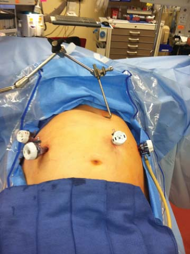
The peritoneal cavity is accessed via a 5-mm port under direct visualization in the left subcostal region using a 5-mm 0° scope. The 5-mm 0° scope is then switched out to a 5-mm 30° scope. Three additional trocars are placed under direct visualization. A 5-mm trocar is placed in the supraumbilical region just left of the midline approximately 18–22 cm from the xiphoid process. A 12-mm trocar is placed on the contralateral side and a 5-mm trocar is placed in the right subcostal margin opposite the initial access trocar for the surgeon’s right and left hand instruments, respectively. A 5-mm incision is also created in the subxiphoid region to allow for the placement of the Nathanson liver retractor (Fig. 6.1). This retractor is used to elevate the lateral segment of the left lobe of the liver and expose the gastroesophageal junction and anterior portion of the stomach.
The gastrocolic omentum is dissected from the stomach permitting entry into the lesser sac. This is performed by cephalad retraction of the greater omentum while incising the avascular plane above the transverse colon (Fig. 6.2). The dissection continues at the pylorus with ligation of the right gastroepiploic artery using a laparoscopic vascular stapler inserted through the 12-mm trocar (Fig. 6.3). This dissection proceeds along the greater curvature of the stomach and ends halfway between the pylorus and the gastroesophageal junction. This maneuver spares the left gastroepiploic vessels and the short gastric vessels. If there is any concern as to the level of division of the stomach to achieve adequate margins an intraoperative endoscopy can help determine the site of gastric resection. The posterior wall of the stomach is separated from the anterior pancreas and base of the transverse mesocolon by blunt dissection and sharp division of connective tissue attachments which can be very inflamed and dense in some patients. The duodenum is carefully kocherized, and in patients with pyloric inflammation, care must be taken to avoid injury to the bowel, common hepatic artery, common bile duct, and portal vein. The right gastric vessels are similarly ligated close to the stomach using a laparoscopic vascular stapling device or ultrasonic shears (Fig. 6.4).
Stay updated, free articles. Join our Telegram channel

Full access? Get Clinical Tree


