Laparoscopic Total Abdominal Colectomy with Ileorectal Anastomosis
KEY STEPS
1. Insertion of ports: 10-mm umbilical Hasson technique; 12-mm right iliac fossa; 5-mm right upper quadrant; 5-mm left upper quadrant; 5- to 10-mm left iliac fossa.
2. Patient rotated to the right and steep Trendelenburg.
3. Greater omentum reflected over the transverse colon, and the small bowel moved to right upper quadrant.
4. Laparoscopic mobilization of the right colon and the terminal ileum.
5. Laparoscopic mobilization of the transverse colon, the middle colic vessels, and takedown of the splenic flexure.
6. Laparoscopic mobilization of the left colon.
7. Mobilization and division of the upper rectum.
8. Confirmation of colonic mobility.
9. Exteriorization of specimen and preservation of mesenteric orientation.
10. Reinsufflation and anastomosis.
11. Closure of ports >5 mm in size
ADDITIONAL ADVICE
1. The correct positioning of the lower limbs—slight flexion of the knee joint and slight extension and abduction of the hip joint—is essential as access to the transverse colon can be impeded if there is any hip flexion.
2. The key elements in the completion of a laparoscopic ileorectal anastomosis are preservation of anastomotic integrity, prevention of torsion of the superior mesenteric arterial supply, and prevention of small bowel internal herniation.
3. Take great care with the middle colic vessels, particularly to avoid bleeding and injury to the pancreas.
4. When mobilizing the left colon, avoid the temptation of making progress into the pelvis, as this is often an easy line of dissection to follow; however, extensive mobilization of the rectum is not indicated in this operation, unless a proctocolectomy is being performed.
5. After reinsufflation, delivering the terminal ileum back into the abdomen on a fixed Allis clamp helps to maintain the alignment of the superior mesenteric artery.
PATIENT POSITIONING
The patient is placed supine on the operating table on a bean bag. After induction of general anesthesia and insertion of an orogastric tube and a Foley catheter, the legs are placed in yellow fin stirrups. The lower aspect of the bean bag should not encroach over the “break” in the operating table and the patient’s buttocks should rest on this edge permitting easy access for a standard circular stapler to be used transanally to complete the ileorectal anastomosis. The arms are tucked at the patient’s side and the bean bag is aspirated. The abdomen is prepared with antiseptic solution and draped routinely (Chapter 2).
INSTRUMENT POSITIONING
The primary monitor is placed on the left side of the patient at approximately the level of the patient’s upper quadrant. The secondary monitor is placed on the right side of the patient at the same level and is primarily for the assistant during the early phase of the operation and port insertion (Fig. 17.1). The operating nurse’s instrument table is placed between the patient’s legs. There should be sufficient space to allow the operator to move from either side of the patient to between the patient’s legs if necessary. The primary operating surgeon stands on the right side of the patient with the assistant standing on the patient’s left, and moving to the right side, caudal to the surgeon once ports have been inserted. A 0-degree camera lens is used.
UMBILICAL PORT INSERTION
This is performed using a modified Hasson approach (Chapter 3). A vertical 1-cm subumbilical incision is made. This is deepened down to the linea alba, which is then grasped on each side of the midline using Kocher clamps. Cautery is used to open the fascia between the Kocher clamps, and Kelly forceps are used to open
the peritoneum bluntly. It is important to keep this opening small (<1 cm) to minimize air leaks. Having confirmed entry into the peritoneal cavity, a purse string of 0 polyglycolic acid is sutured around the subumbilical fascial defect (umbilical port site) and a Rommel tourniquet is applied. A 10-mm reusable port is inserted through this port site allowing the abdomen to be insufflated with CO2 to a pressure of 12 mmHg.
the peritoneum bluntly. It is important to keep this opening small (<1 cm) to minimize air leaks. Having confirmed entry into the peritoneal cavity, a purse string of 0 polyglycolic acid is sutured around the subumbilical fascial defect (umbilical port site) and a Rommel tourniquet is applied. A 10-mm reusable port is inserted through this port site allowing the abdomen to be insufflated with CO2 to a pressure of 12 mmHg.
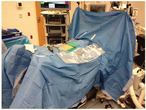 FIGURE 17.1. Room setup. |
LAPAROSCOPY AND INSERTION OF REMAINING PORTS
The camera is inserted into the abdomen and an initial laparoscopy is performed carefully evaluating the liver, small bowel, and peritoneal surfaces. A 12-mm port is inserted in the right lower quadrant approximately 6 to 8 cm medial and superior to the anterior superior iliac spine. In patients marked for a temporary ileostomy, the port is placed through this site, paying careful laparoscopic attention to the position of the inferior epigastric vessels to avoid injury. A 5-mm left lower quadrant port is used in order to permit use of an energy source when ligating the ileocolic pedicle and middle colic vessels. This port site may be upsized to 10 mm in size as needed to accommodate the energy sources. Five-millimeter ports are then inserted in the right upper quadrant and left upper quadrant at least a hand’s breadth superior to the lower quadrant ports. The left-sided ports are kept lateral to the epigastric vessels (Fig. 17.2).
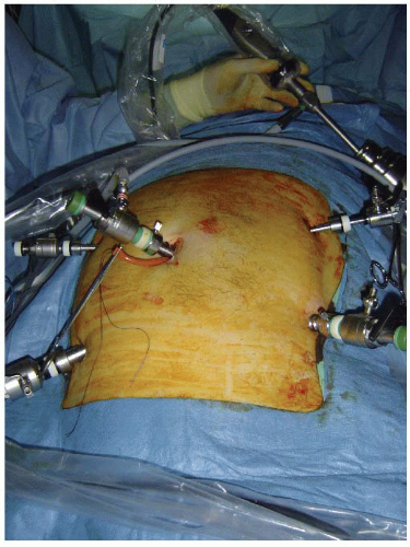 FIGURE 17.2. Port insertion for a total abdominal colectomy (a 12-mm port in the right lower quadrant, 5-mm ports in the right upper quadrant, left lower quadrant, and left upper quadrant). |
DEFINITIVE LAPAROSCOPIC SETUP
The assistant now moves to the patient’s left side, standing caudad to the surgeon. The patient is rotated with the right side up and left side down, to approximately 15 to 20 degrees tilt, and often as far as the table can go. This helps to move the small bowel over to the left side of the abdomen. The patient is then placed into the Trendelenburg position. This again helps gravitational migration of the small bowel away from the operative field. The surgeon then inserts two atraumatic bowel clamps through the two left-sided abdominal ports. The greater omentum is reflected over the transverse colon so that it comes to lie on the stomach. If there is no space in the upper part of the abdomen, one must confirm that the orogastric tube is adequately decompressing the stomach of gas. The small bowel is moved to the patient’s left side, some remaining in the pelvis and the upper abdomen, allowing visualization of the ileocolic pedicle. This may necessitate the use of the assistant’s 5-mm atraumatic bowel clamp through the right lower quadrant port in order to tent the ileal mesentery medially and cephalad.
LAPAROSCOPIC MOBILIZATION OF THE RIGHT COLON AND THE TERMINAL ILEUM
The surgeon initially uses two bowel graspers to expose the ileocolic pedicle. The assistant standing opposite the surgeon using a 5-mm bowel grasper secures the ileocecal junction and elevates this laterally and toward the right lower quadrant. This has the effect of the ileocolic vascular pedicle been drawn up from the retroperitoneum demonstrating the line of the vessels (Fig. 17.3). The surgeon uses a bowel grasper in his left hand and electrocautery in his right hand and incises along the line of the exposed vessels. This blunt dissection continues using traction and counter-traction opening up the underside of the ascending colonic mesentery laterally and exposing the second part of the duodenum medially. The optimal movement is generally to elevate the ascending colon mesentery with the left hand instrument and dissect or separate tissues off this with the right hand instrument.
The ileocolic pedicle should be exposed and a potential avascular window appears between the superior/lateral aspect of the base of the ileocolic pedicle and the duodenum. The surgeon now uses the bowel grasper to elevate this avascular window from the superior aspect (superficial) and using sharp dissection incises through into the virtual space opened up on the underside of the ascending colonic mesentery. The surgeon now places his scissors under the ileocolic pedicle and out through this newly created window and confirms its suitability for ligation. The ileocolic pedicle is then divided (Fig. 17.4). This technique is outlined in detail in Chapter 5. The assistant now drops the ileocecal junction and grasps the distal end of the transected ileocolic pedicle. The assistant elevates this along with the mesentery and the surgeon progressively dissects under the ascending mesentery to reach the lateral abdominal wall, the lower aspect of the liver and the underside of the transverse colonic mesentery. It is important to dissect as far medially under the transverse colon while avoiding any
duodenal or pancreatic injury. A low rising right colic artery, or right branch of middle colic vessels, may be encountered while dissecting cephalad, and this should be ligated with surgical clips, or an energy source.
duodenal or pancreatic injury. A low rising right colic artery, or right branch of middle colic vessels, may be encountered while dissecting cephalad, and this should be ligated with surgical clips, or an energy source.
Related posts:


Stay updated, free articles. Join our Telegram channel

Full access? Get Clinical Tree


