Laparoscopic Right Hemicolectomy
KEY STEPS
1. Insertion of ports: 10-mm umbilical Hasson technique; 5-mm left iliac fossa; 5-mm left upper quadrant; 5-mm right iliac fossa (optional).
2. Patient rotated fully to the left and slightly Trendelenburg.
3. Laparoscopic assessment and then the small bowel and omentum are moved toward the left upper quadrant.
4. Ileocolic pedicle defined and divided, protecting the ureter and the duodenum.
5. Full medial-to-lateral mobilization of the cecum, ascending colon, and hepatic flexure.
6. Division of right branch of the middle colic vessels.
7. Hepatic flexure mobilization completed using a superior approach.
8. Cecum retracted up and laterally to complete mobilization of the cecum and the small bowel mesentery off the retroperitoneum.
9. Confirmation of full mobilization of the right colon to the midline.
10. Extension of umbilical port site and exteriorization of the right colon.
11. Standard extracorporeal resection and anastomosis.
ADDITIONAL ADVICE
1. When performing medial mobilization of the ileocolic artery for cancer, make your initial peritoneal incision close to the SMA to ensure a complete mesocolic excision.
2. As the ileocolic pedicle is mobilized, Toldt’s fascia is carefully protected on the retroperitoneum, thereby protecting the ureter, the duodenum, and the retroperitoneal structures.
3. When performing the medial-to-lateral mobilization of the ascending colon mesentery, draw the mesentery medially and anteriorly to stretch out the areolar attachments, facilitating your dissection.
4. Be sure to extend your medial mobilization inferiorly behind the cecum and superiorly over the duodenum and head of pancreas right up to the lower
border of the liver. Sometimes, one even opens through the hepatico-colic ligament using this approach.
border of the liver. Sometimes, one even opens through the hepatico-colic ligament using this approach.
5. It is often easier to leave division of the right branch of the middle colic artery until after full mobilization of the hepatic flexure, especially in more obese individuals.
6. When performing the superior part of the hepatic flexure mobilization, draw the colon anteriorly and inferiorly to maximize the space behind the hepatic flexure which has already been dissected.
7. For patients with a cancer near the hepatic flexure or transverse colon, the greater omentum is taken en bloc with the colon.
8. Mobilizing the greater omentum and transverse colon to or beyond the midline facilitates specimen extraction, particularly in more obese individuals.
9. In rare cases for extremely obese patients, a right upper quadrant port can be used. In these very obese patients, placing the “left-sided” ports through the midline may also help to reach the right upper quadrant.
PATIENT POSITIONING
The patient is placed supine on the operating room (OR) table on a bean bag. After induction of general anesthesia and insertion of an oral gastric tube and Foley catheter, the legs are placed in yellow fin stirrups. The arms are tucked at the patient’s side and the bean bag is aspirated. For more obese patients who will not fit on the OR table, the right arm is left out from the side. The abdomen is prepared with antiseptic solution and draped routinely.
INSTRUMENT POSITIONING
The primary monitor is placed on the right side of the patient at about the level of the shoulder. The secondary monitor is placed on the left side of the patient at the same level and is primarily for the assistant during the early phase of the operation and port insertion (see Fig. 12.1). The operating nurse’s instrument table is placed between the patient’s legs. The primary operating surgeon stands on the left side of the patient with the assistant standing on the patient’s right, and moving to the left side, caudad to the surgeon once ports have been inserted. A 0-degree camera lens is preferred.
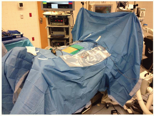 FIGURE 12.1. Room setup for right hemicoloectomy. |
UMBILICAL PORT INSERTION
This is performed using a modified Hasson approach (Chapter 3). A vertical 1-cm subumbilical incision is made. This is deepened down to the linea alba, which is then grasped on each side of the midline using Kocher clamps. Cautery is used to open the fascia between the Kocher clamps and Kelly forceps are used to open the peritoneum bluntly. It is important to keep this opening small (1 cm) to minimize air leaks. Having confirmed entry into the peritoneal cavity, a purse string of 0 polyglycolic acid is sutured around the subumbilical fascial defect (umbilical port site) and a Rommel tourniquet applied. A 10-mm reusable port is inserted through this port site allowing the abdomen to be insufflated with CO2 to a pressure of 12 to 15 mmHg.
LAPAROSCOPY AND INSERTION OF REMAINING PORTS
The camera is inserted into the abdomen and an initial laparoscopy is performed, carefully evaluating the liver, small bowel, and peritoneal surfaces. A 5-mm port is inserted in the left lower quadrant approximately 2 to 3 cm medial and superior to the anterior superior iliac spine. This is carefully inserted lateral to the inferior epigastric vessels, paying attention to keep the tract of the port going as perpendicular
as possible through the abdominal wall. A 5-mm port is then inserted in the left upper quadrant at least a hand’s breadth superior to the lower quadrant port. Particularly when teaching, a 5-mm right lower quadrant port is also inserted. Rarely, in the case of a difficult hepatic flexure, a 5-mm right upper quadrant port may also be needed (Fig. 12.2).
as possible through the abdominal wall. A 5-mm port is then inserted in the left upper quadrant at least a hand’s breadth superior to the lower quadrant port. Particularly when teaching, a 5-mm right lower quadrant port is also inserted. Rarely, in the case of a difficult hepatic flexure, a 5-mm right upper quadrant port may also be needed (Fig. 12.2).
Related posts:


Stay updated, free articles. Join our Telegram channel

Full access? Get Clinical Tree


