Laparoscopic Revision of Failed Fundoplication and Hiatal Hernia

Chapter 7 Laparoscopic Revision of Failed Fundoplication and Hiatal Hernia


The videos associated with this chapter are listed in the Video Contents and can be found on the accompanying DVDs and on Expertconsult.com.image


Laparoscopic fundoplication has been embraced by the surgical community as the procedure of choice for gastroesophageal reflux disease (GERD). After the introduction of laparoscopy in foregut surgery, a significant rise in the number of laparoscopic fundoplications allowed for evaluation of long-term outcomes in large patient series. Hiatal hernia recurrence has been shown to be a significant factor in the failure of antireflux procedures. Patients with failed fundoplication often suffer from persistent, recurrent, or new-onset symptoms.


Redo fundoplication represents one of the most technically challenging procedures in laparoscopic surgery; in addition, complex clinical and diagnostic logistics come into play. Invariably in these cases, the laparoscopic surgeon encounters distorted anatomy, dense adhesions, and fibrotic tissue in proximity to structures such as the esophagus, aorta, liver, and spleen. Success of revisional operations depends on the primary procedure, the patient’s symptoms, the results of preoperative tests, and, more important, patient selection.


In this chapter, the absolute and relative indications for revisional fundoplication and hiatal herniorrhaphy and the value of preoperative examinations in selecting the appropriate medical or surgical treatment are discussed. Furthermore, the technique of revisional surgery for failed fundoplication and failed hiatal hernia repair is described, with emphasis on operative risks and pitfalls. Finally, the morbidity and the outcomes of the procedure are outlined.


Operative indications


Patients with “failed fundoplication” may be divided into those with anatomic failure and those with a normal anatomy but persistent foregut symptoms. The latter are subdivided into patients with objective evidence of gastroesophageal reflux, esophageal stenosis, or delayed gastric emptying, and patients with no functional, endoscopic, or imaging evidence that could explain their symptoms.


Anatomic Failure


Seven types of anatomic failure may be encountered.


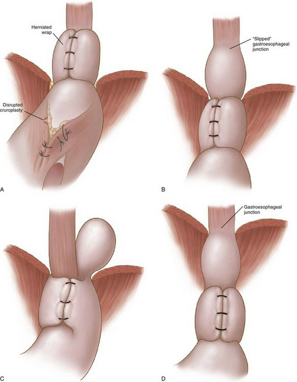
Wrap migration (44%). A portion of the wrap or the entire wrap has migrated into the mediastinum (Fig. 7-1A). The proposed etiologic factor is inadequate crural closure owing to poor technique or weak tissue, or both; disruption of the cruroplasty results in migration of the wrap into the mediastinum.


Slipped hernia (16%). The gastroesophageal junction has slipped into the mediastinum, whereas the wrap remains below the diaphragm (Fig. 7-1B). It has been postulated to result from the presence of a short esophagus or inadequate mobilization of the esophagus during the primary procedure. The latter is a more probable mechanism.


Paraesophageal hernia (16%). Both the body of the wrap and the gastroesophageal junction remain below the diaphragm, whereas a part of the stomach has migrated into the mediastinum, posterior to the esophagus (Fig. 7-1C). This type of failure is a result of the combination of a loose wrap and a poor crural closure, which allows part of the posterior portion of the wrap to migrate into the mediastinum.


Displaced wrap (10%). The wrap has slipped caudad on the body of the stomach, resulting in formation of a fundal pouch (Fig. 7-1D). The reasons for this type of failure are elusive; inadequate anchorage of the fundoplication may play a role. Retention of food in the herniated pouch may result in an increase of its size and further caudad slippage of the wrap (hourglass stomach).


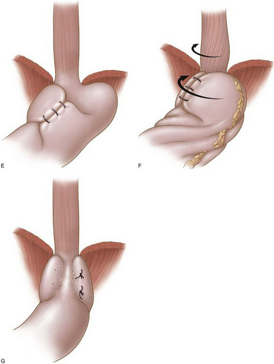
Misplaced wrap (4%). The plication has been constructed by the fundus and the body of the stomach because of misidentification of the anatomy at the initial operation (Fig. 7-1E).


Twisted wrap (6%). Torsion of the wrap is thought to result from insufficient division of the short gastric vessels and continuous traction counterclockwise (Fig. 7-1F).


Disrupted wrap (4%). A part of the fundoplication or the entire fundoplication has been disrupted. Inadequate construction of the fundoplication (superficially placed sutures) may account for this failure (Fig. 7-1G).


Recurrence usually occurs either during the early postoperative period or in the first 2 years after surgery. Wrap migration is the most common cause for failure. Excessive retching, nausea, and vomiting have been identified as causative factors for failure within the first 2 postoperative weeks; therefore, routine administration of antiemetics after laparoscopic fundoplication has been advocated by many. Early hernia recurrence is considered an indication for revisional fundoplication.


Late fundoplication failure may present with or without foregut symptoms. The management of symptomatic patients with documented failed fundoplication can be either conservative or operative. Revisional surgery is an effective treatment for individuals with hiatal hernia recurrence and reflux symptoms. Conservative treatment may be considered for high-risk patients or for those with multiple previous abdominal operations. However, patients presenting with hiatal hernia recurrence and dysphagia or gas-bloat syndrome are difficult to manage conservatively.


Preoperative evaluation, testing, and preparation


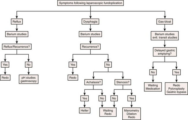
A thorough clinical examination and a detailed clinical history are essential for patients with suspected failed fundoplication. Other pathologies of the upper gastrointestinal tract, such as gastritis, peptic ulcer, pancreatitis, cholelithiasis, and cardiac and pulmonary diseases, should be ruled out. Presenting symptoms and the findings of functional and imaging studies should be reviewed carefully. Selective use of barium studies, upper gastrointestinal endoscopy, gastric emptying studies, esophageal pH monitoring, and manometry will determine the form of treatment.


< div class='tao-gold-member'>

Stay updated, free articles. Join our Telegram channel

Jul 20, 2016 | Posted by in GASTOINESTINAL SURGERY | Comments Off on Laparoscopic Revision of Failed Fundoplication and Hiatal Hernia

Full access? Get Clinical Tree

Get Clinical Tree app for offline access