Laparoscopic Radical Cholecystectomy

Chapter 15 Laparoscopic Radical Cholecystectomy


The videos associated with this chapter are listed in the Video Contents and can be found on the accompanying DVDs and on Expertconsult.com.image


Gallbladder cancer is a disease process that has a diverse worldwide variation. It has a very high incidence in parts of northern India, Pakistan, Bolivia, Peru, and Ecuador. It also has a notable presence in South Asia and some central and eastern European countries. Females have a higher incidence, with factors such as cholelithiasis, obesity, and infections related to Salmonella paratyphi and typhi showing a higher relative risk. It has been estimated that approximately 1% of patients with gallstones in Western countries will develop gallbladder cancer. In areas of high risk such as Ecuador, this increases to 5% to 10% and may be as high as 20% in Bolivia and Peru. A subset of patients with cholelithiasis will develop porcelain gallbladder, which is a consequence of a chronically inflamed wall. However, not every patient with a porcelain gallbladder will develop gallbladder cancer, with the risk more in the range of 10% to 20%. Despite these possible etiologic factors, the exact pathway behind gallbladder cancer remains veiled and likely multifactorial.


As an anatomic structure, the gallbladder lies below hepatic segments IVB and V with close proximity to the portal structures. Because it has only one muscle layer, the tumor has easier access to the serosa of adjacent organs, and its close proximity to the structures of the hepatoduodenal ligament often make surgical resection difficult or impossible. Of note, the gallbladder has an adventitial layer along its attachment to the liver and a serosa only along its extrahepatic portion. The first-echelon nodes of drainage are the cystic and pericholedochal nodes, with connection further to portal and common hepatic artery nodes, making their dissection a critical part of any surgical resection. The disease process often frustrates because of delays in diagnosis, resulting in presentation at an advanced stage and incurability. For those who present at an earlier stage, surgery remains the only chance of cure, with the consideration of postoperative chemotherapy and radiation.


In the era of laparoscopic cholecystectomy, the incidental identification of gallbladder cancer represents the majority of presentations for this disease process. Gallbladder cancer is a relatively rare disease, with the incidence rate in the United States estimated to be approximately 1.2 cases per 100,000 per year. It is associated with a poor prognosis, with 5-year survival rates for gallbladder cancer being reported at 5% to 10% in recent years and with a median survival of 3 to 6 months from the time of diagnosis. This has been improving, however, with groups reporting median survival of 50 months for those amenable to surgical resection. Since George Pack first suggested in 1955 a radical liver resection for gallbladder cancer, there is consideration of partial hepatectomy ranging from wedge resection to formal hepatectomy. The advent of laparoscopy to the field of hepatobiliary surgery has further added to the surgical approach and bears discussion.


Advent of the laparoscopic approach


Surgeons have been reluctant to apply minimally invasive techniques to the surgical management of gallbladder cancer because of the perceived difficulty of laparoscopically dissecting tumors off the structures of the portal triad. As a result, laparoscopic radical cholecystectomy has been one of the last minimally invasive procedures performed. Gumbs and colleagues reported the first laparoscopic radical cholecystectomy in 2009 on a patient with the preoperative suspicion of gallbladder cancer due to a 4-cm mass seen on abdominal ultrasound and cross-sectional imaging. Despite a negative preoperative serum immunoglobulin G4 (IgG4) level, the postoperative diagnosis was consistent with autoimmune cholecystitis. Nonetheless, the patient was discharged home tolerating a regular diet on the second postoperative day.


The next patient with the preoperative suspicion of gallbladder cancer was found to have a 7.5-cm malignant mass in the dome of the gallbladder, with intraparenchymal invasion during the laparoscopic radical cholecystectomy; as a result, a formal resection of hepatic segments IVB and V was required to obtain an R0 resection. In addition, the cystic duct stump at the confluence with the common bile duct was found to have disease. A laparoscopic common bile duct excision was then performed and the biliary tree reconstructed with a laparoscopic Roux-en-Y choledochojejunostomy. Because of the success with these patients, we began to approach patients who had undergone previous cholecystectomy and were found to have gallbladder cancers.


Postoperatively diagnosed gallbladder cancer


The need for re-resection following an incidentally diagnosed gallbladder cancer depends on the final pathologic stage of the tumor. By definition, T1a tumors only invade the lamina propria. A cholecystectomy with negative margins done laparoscopically or by open techniques is considered curative because re-resection of the gallbladder fossa with or without lymphadenectomy has never been proved to yield a survival benefit. Re-resection, however, is advised in patients with T1b to T3 tumors because of improved overall survival. Patients with incidentally diagnosed gallbladder cancer often have a better prognosis because most are early lesions (T1 to T2). Patients found to have incidental gallbladder carcinomas who undergo re-resection may actually have improved survival compared with those with nonincidentally diagnosed cancer. T2 lesions make up the majority (67%) of incidentally diagnosed gallbladder carcinomas and are associated with a 5-year survival rate of greater than 60%, compared with less than 20% for those treated with cholecystectomy alone. Patients with T3 disease, which is defined as a tumor that invades into the serosa and/or the liver and/or an adjacent organ, are at a higher risk for peritoneal carcinomatosis. Formal re-resection and hepatoduodenal lymphadenectomy may provide a survival benefit in patients without evidence of peritoneal disease. Patients with improperly treated T3 disease have a 5-year survival rate of 0 to 15% compared with 25% to 65% for T3 patients who undergo completion radical cholecystectomy re-resection. The extend of liver resection for gallbladder malignancy is still ill defined.


Because of the authors’ success with these patients and the ability to spare patients unnecessary laparotomy, all resectable patients with preoperatively suspected gallbladder cancer and patients found to have T1b to T3 disease after cholecystectomy are now approached laparoscopically.


Operative indications


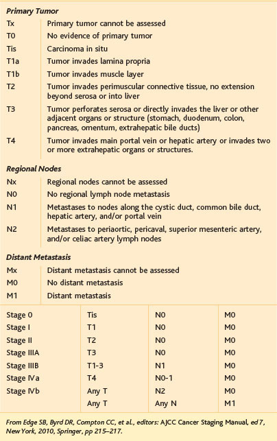
Operability in gallbladder cancer relies heavily on preoperative staging and considerations of findings during the initial laparoscopy or as part of a preoperative staging workup. The American Joint Committee on Cancer (AJCC) in its seventh edition has established subcategories for stage IV gallbladder cancer. Stage IVA is defined by tumor invasion into the main portal vein, the hepatic artery, or two or more extrahepatic structures. The presence of metastases to the periaortic, pericaval, or superior mesenteric artery or celiac nodes or of distant metastases further defines stage IVB (Table 15-1). As mentioned previously, the extent of liver resection needs strong consideration and is controversial, with differing opinions regarding the suitability of nonanatomic resections of the gallbladder bed versus complete resection of segments IVB and V and even extended right hepatectomy (segments IV to VIII). We consider major hepatectomy or even extended hepatectomy only when this is necessary to achieve an R0 resection because there has been no conclusive difference noted in overall survival between minor and major hepatectomy.


Nodal disease stretching down the portal chain posterior to the pancreas or duodenum may present a challenge. For disease that is not invading vascular structures, dissection is often achieved satisfactorily; however, larger nodes may not be removed successfully without a pancreatoduodenectomy. This must be taken with due caution in terms of morbidity and mortality when performing such a procedure allied with a radical cholecystectomy.


Powered analysis suggests that involvement of the common bile duct is associated with advanced T stage and is an independent prognostic factor in survival. Positive margins at the cystic duct mandate further resection of the common bile duct to achieve R0 resection, which, although a poor prognosticator, is associated with a better survival outcome than R1 resection.


Preoperative evaluation


Routine preoperative workup includes chest radiography, electrocardiography (ECG), serum complete blood count, chemistries, liver function studies, and serum tumor markers consisting of carcinoembryonic antigen and CA 19-9 levels. In addition, serum IgG4 levels are measured routinely in all patients before surgery to rule out the presence of autoimmune cholecystitis. Unfortunately, as noted previously, this test has a significant false-positive rate. The indocyanine green test is routine in Asia because the high rate of hepatitis-related cirrhosis may influence the extent of hepatic resection. All formal hepatectomies must be assessed for residual liver volume and Child-Pugh classification and should be followed by pathologic examination for underlying liver cirrhosis grade, especially if the liver is not chemotherapy naïve.


Ultrasound is limited in the diagnosis of early lesions and as such is unreliable for staging. In some 20% to 30% of cases, gallbladder cancer may present as an asymmetrical wall thickening that has an expanded differential diagnosis ranging from cholecystitis, adenomyomatosis, acute hepatitis, or portal hypertension to congestive heart failure. Gallbladder cancer arising on a background of chronic inflammation certainly makes radiologic interpretation more difficult. Asymmetrical wall thickening with persistent arterial enhancement or isodensity during the hepatic venous phase should, however, heighten suspicion. In cases in which a mass-occupying lesion is noted, as occurs in some 40% of patients, ultrasound that shows a heterogenous and hypoechoic tumor is classic.


Although ultrasound is often the first imaging technique performed in those undergoing cholecystectomy for unsuspected gallbladder cancer, high-resolution imaging in the form of computed tomography (CT) or magnetic resonance imaging (MRI) is most often used to help identify residual tumor burden or metastatic disease. CT scan with intravenous contrast is used to delineate the hepatic vasculature and rule out evidence of vascular invasion, such as portal vein or hepatic arterial narrowing or frank invasion. Abdominal MRI with gadolinium is used in an effort to ascertain the extent of intraparenchymal involvement. In cases of recent cholecystectomy, it may be difficult or impossible to differentiate hepatic invasion from enhancement because of scarring from the recent surgery.


The role of [18]F-fluorodeoxyglucose positron emission tomography (FDG-PET) in gallbladder cancer is still in flux. Because gallbladder cancer is highly PET avid, studies have suggested that FDG-PET may change the operative decision in 25% of cases. In up to 50% of cases of incidentally found gallbladder cancer, metastatic spread was shown by FDG-PET. Although this is highly suggestive of a role for FDG-PET, false-positive results may be noted in areas of inflammation from recent laparoscopic cholecystectomy, with recent data also noting FDG-PET to have a negative predictive value of 65%, suggesting a greater extent of residual disease that might be missed. It may be that the role of FDG-PET is best served to rule out distant spread, whereas residual disease is best assessed by intraoperative laparoscopic ultrasound and reexploration.

< div class='tao-gold-member'>

Stay updated, free articles. Join our Telegram channel

Jul 20, 2016 | Posted by in GASTOINESTINAL SURGERY | Comments Off on Laparoscopic Radical Cholecystectomy

Full access? Get Clinical Tree

Get Clinical Tree app for offline access