Laparoscopic Partial Nephrectomy

Chapter 32 Laparoscopic Partial Nephrectomy


imageThe videos associated with this chapter are listed in the Video Contents and can be found on the accompanying DVDs and on Expertconsult.com.


Relative to radical nephrectomy (RN), nephron-sparing surgery with partial nephrectomy (PN) has been demonstrated to provide equivalent oncologic control, improved renal function, a lower risk for cardiovascular disease, and improved overall survival. Techniques developed for obligatory PN (solitary kidney, renal insufficiency, synchronous tumors, genetic predisposition) have also been used in the setting of a healthy contralateral renal unit. As a consequence, RN is no longer an acceptable option when a PN is indicated and is technically feasible. Moreover, wide dissemination of surgical techniques and continued improvement in instrumentation have made PN the preferred approach for small masses of the kidney.


More recently, laparoscopic partial nephrectomy (LPN) has been shown to have functional and oncologic outcomes comparable to those of open surgery. LPN is technically more demanding to perform, and it has been shown to have a steep learning curve. In this chapter, we describe our transperitoneal technique for LPN and discuss strategies for the prevention and management of complications.


Operative indications


The indications for partial nephrectomy include bilateral tumors or a mass in the presence of renal insufficiency or in a solitary functioning kidney. In addition, the indications for PN now include the patient with a normal contralateral kidney. The indications for LPN have broadened to include more challenging cases, such as tumors larger than 4 cm, multiple tumors, hilar tumors, and completely endophytic masses.


Contraindications for LPN are similar to those for open PN, including active peritonitis, coagulopathy, bowel obstruction, and severe cardiopulmonary disease. Renal anatomic anomalies, such as horseshoe or ectopic kidney, do not preclude the laparoscopic approach, provided that adequate preoperative imaging has been obtained to delineate renal vascular supply. Relative contraindications are related to the surgeon’s experience and comfort level. A history of previous abdominal or ipsilateral kidney surgery may be considered a relative contraindication. A retroperitoneal approach may be preferable in such a case to avoid intra-abdominal adhesions.


Obesity had been considered a relative contraindication to LPN. Initial access in obese patients (body mass index [BMI] >30 kg/m2) can be difficult, resulting in a higher likelihood of abdominal wall vessel injury and subcutaneous dissection. Longer trocars and bariatric laparoscopic instrumentation should be available. Skin-to-peritoneum distance can be calculated from abdominal imaging. Port placement and entry angle to the subcutaneous tissue should allow for instrument triangulation and freedom of movement. With adequate operative experience, LPN in obese patients may be attempted. The Cleveland Clinic experience with obese patients (BMI >30 kg/m2) has suggested that LPN is technically feasible in this patient group. Specifically, when compared with a cohort of open PN controls, obese patients undergoing LPN experienced reduced blood loss and analgesic requirement, quicker return of bowel function, reduced hospital stay, and shorter convalescence; operative times and complication were similar. Comparing obese and nonobese subjects undergoing LPN, other authors have reported longer operative times and increased blood loss with the obese group, but with similar complication and conversion rates and recovery period. Overall, an obese patient appears to have a comparable outcome to that of a nonobese patient when undergoing LPN by an experienced surgeon. We recommend that initial operative experience with LPN should be obtained with nonobese patients to minimize the risk for complications and open conversion.


Although PN remains the standard of care for small renal masses, ablative techniques have demonstrated acceptable oncologic outcomes in series with short-term follow-up, and represent viable alternatives to PN. Preliminary studies appear to have favored cryoablation rather than radiofrequency ablation, but this may change with additional data. Interestingly, observational series (i.e., no resection or ablation) have shown that very few patients demonstrate disease progression while on active surveillance. Some have suggested that delayed intervention may be a viable management strategy.


Preoperative evaluation, testing, and preparation


As part of the preoperative workup, all patients should have a complete history and physical examination, with particular attention to prior abdominal surgical history. During the assessment, the patient’s body habitus, location of previous surgical incisions, and presence of skeletal deformities should be noted. Each of these factors can influence the choice of laparoscopic approach, patient positioning, and port placement. The informed consent discussion should include the risk for bleeding, injury to peritoneal contents, and the possibility of open conversion, which occurs in about 5% of LPN procedures.


Preoperative laboratory studies include a complete blood count, serum chemistries, coagulation panel, urinalysis, and urine culture. Chest radiograph and electrocardiogram also are obtained. Additional pulmonary function studies and cardiac workup are ordered as needed. Clinical suspicion of advanced disease or metastasis should entail additional imaging, such as a chest computed tomography (CT), head CT, and bone scan. A type and screen is obtained, and two units of packed red blood cells are crossmatched. Anticoagulants and platelet inhibitors should be stopped 5 to 7 days before surgery.


Candidates for LPN should undergo a staging workup with CT or magnetic resonance imaging (MRI). An abdominopelvic CT scan, with and without intravenous contrast and with a 10-minute delayed phase and coronal and sagittal reconstructions, is our imaging modality of choice. This study provides an excellent depiction of perirenal anatomy, including the location and number of renal vessels, as well as the location of the ureter. A CT scan is helpful in detecting the presence of aberrant renal vessels, which are known to occur in 25% to 40% of kidneys. If an accessory vessel is suspected but not well defined on CT, some surgeons recommend that an angiogram (CT, MRI, or conventional) should be performed to rule out the presence of vascular variants.


CT also allows the surgeon to assess the relationship of the kidney (position and rotation) to adjacent structures and to obtain an accurate estimate of the amount of perirenal fat present. In patients with a history of atherosclerosis, one should carefully examine the noncontrasted images of the CT scan to identify renal artery wall calcifications. If mural calcification is present, particularly at the ostium, the renal artery should be dissected and clipped at a point where the vessel is free of atherosclerotic disease. Vessel fracture during clip application can cause severe arterial hemorrhage. Imaging also can be used to calculate the RENAL (radius of tumor; exophytic/endophytic properties, nearness of tumor to the collecting system; anterior/posterior descriptor; location relative to the polar line) Nephrometry Score (RNS), a standardized classification system that quantifies the salient anatomy of renal masses. In patients undergoing LPN, a higher RNS is associated with an increased estimated blood loss, warm ischemia time, and length of hospital stay.


Gadolinium-enhanced MRI also is a suitable consideration for patients with renal impairment or contrast allergy. Finally, if there are concerns regarding the residual renal function, a MAG-3 (mercaptoacetyl triglycine, chelated to technetium-99m) nuclear scan with differential function may be useful.


Preoperative bowel preparation rarely is needed, and depends on the anticipated difficulty of the case. In our experience, if the kidney is not involved with an inflammatory process, then the patient is placed on a clear liquid diet the day before surgery and the bowel preparation omitted. Another option is a limited bowel preparation protocol consisting of a clear liquid diet and a bottle of magnesium citrate the day before surgery. If significant difficulty with dissection is expected, then the patient should undergo a full mechanical bowel preparation along with antibiotics the day before surgery. All patients should be given a parenteral antibiotic, usually a first-generation cephalosporin, in the preoperative holding area. Subcutaneous heparin and pneumatic compression stocking are administered before anesthesia induction. An orogastric tube and Foley catheter also are inserted before patient positioning.


Patient positioning in the operating suite


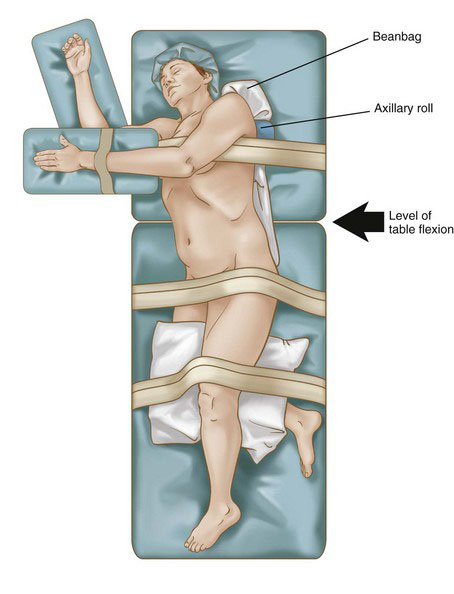
The patient is placed in a modified lateral decubitus position using an underlying beanbag, with the thorax rotated back slightly at 30 degrees (Fig. 32-1). The lower hand is padded and placed on an armrest. The lower leg is flexed at the knee 90 degrees, and the upper leg is left extended. Pillows are placed between the legs for adequate support. Padding is placed under the lower ankle to relieve pressure in this area. An axillary roll then is placed 5 cm caudal to the axilla to protect the brachial plexus from a stretch injury. Additional padding is placed under the lower elbow to prevent ulnar nerve compression. The beanbag then is placed on vacuum to secure the final position. The umbilicus and spine should be visible to ensure adequate exposure in the rare need for an emergent open conversion. Surgical towels are placed over the skin at the hip and knee levels, and 3-inch silk tape is wrapped circumferentially at these levels to completely secure the patient to the table. Finally, an armrest is fastened to the table and secured to support the padded ipsilateral arm. Careful attention to final position and pressure padding is essential because bed rotation during surgery is often required to optimize exposure and assist with gravity bowel retraction. Care is taken not to obstruct any intravenous line.


Stay updated, free articles. Join our Telegram channel

Jul 20, 2016 | Posted by in GASTOINESTINAL SURGERY | Comments Off on Laparoscopic Partial Nephrectomy

Full access? Get Clinical Tree

Get Clinical Tree app for offline access