Laparoscopic Myomectomy

Chapter 37 Laparoscopic Myomectomy


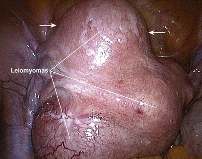
A myoma is a benign solid tumor consisting of fibrous tissue; hence, it often is called a fibroid tumor. A uterine leiomyoma, or fibroid, refers to a fibrous uterine tumor of smooth muscle origin. A leiomyoma is a well-circumscribed, round solid tumor, pearly white or light tan in color, that may grow as a single nodule or in clusters of varying size. This tumor may be attached to or be within the myometrium and is pseudoencapsulated by fibrous connective tissue. The diameter of a leiomyoma may range from 1 mm to more than 30 cm (Fig. 37-1).


The cumulative risk for the diagnosis of uterine leiomyoma in the age range of 25 to 44 years is about 30 percent. It is not clear whether leiomyoma is a precursor for leiomyosarcoma, the malignant phenotype. The incidence of leiomyosarcoma is extremely low in premenopausal compared with postmenopausal women, in whom leiomyosarcoma accounts for about 1% of uterine malignancies. The cellular morphology of a leiomyosarcoma is similar to normal myometrial smooth muscle cells.


The pathoetiology of leiomyomatous disease has not been determined, but most fibroids develop in women during their reproductive years. A leiomyoma will not develop before the body begins estrogen production; during pregnancy, a leiomyoma may grow quickly because of elevated estrogen levels. Once menopause has been reached, a leiomyoma generally will stop growing, and even can begin to shrink.


Most fibroids are slow growing, cause no symptoms, and do not need to be treated. About 25% of leiomyomas will cause symptoms, however, and may need medical or surgical treatment. Symptoms can include prolonged or heavy menstrual bleeding, anemia, mass effect, low back or pelvic pressure or pain, urinary frequency, constipation, dyspareunia, and in rare cases, reproductive dysfunction. Resection is the most effective treatment for leiomyomatous disease, and this has been the most common indication for hysterectomy.


In addition to hysterectomy and abdominal myomectomy, laparoscopic myomectomy has been developed to treat leiomyomatous disease. Kurt Semm reported the first laparoscopic myomectomy in 1979. This procedure is not without controversy, especially with regard to the volume and number of the myomas that can be removed and the putative risk on a subsequent pregnancy. In this chapter, we discuss the indications, technique, outcome, and controversies of laparoscopic myomectomy for uterine leiomyomatous disease.


Operative indications


Traditionally, symptomatic fibroids have been treated with transabdominal myomectomy or hysterectomy. Hysterectomy is the most common major gynecologic operation in the world, and leiomyomatous disease is the most common cause of hysterectomy. For the patient with symptomatic fibroids who would like to retain her uterus, myomectomy should be considered. The indications for myomectomy include fibroid-associated infertility, tumor enlargement, pelvic pain, and abnormal uterine bleeding. In addition, there needs to be at least one myoma 4 cm or larger that is not accessible by hysteroscopy. Submucosal leiomyomas may be removed by hysteroscopy.


No upper size limit for fibroid resectability exists as long as the myoma can be mobilized. The maximal number of myomas that can be resected is somewhat controversial; some authors suggest no more than three or four myomas with a diameter less than 7 to 8 cm; others suggest eight or more myomas, whereas others suggest that the maximal resection number be based on individual choice, pathologic findings, and surgical expertise. Our experience has indicated that the upper limits of size and number for uterine leiomyoma resection should depend on tumor location, depth of penetration, and ease of mobilization rather than the total number of tumors or the maximal diameter. Nevertheless, it should be recognized that previous studies have indicated that the recurrence and complication rates are higher in cases of myomectomy for multiple fibroids.


If the quantity of myomas favors performance of a hysterectomy, then there are a number of less invasive approaches that can be offered to the patient, such as: (1) laparoscopic or percutaneous cryoablation; (2) laparoscopic or percutaneous myolysis with a variety of energy sources; (3) uterine artery embolization (UAE); or (4) magnetic resonance imaging (MRI)-guided focused ultrasound. All these procedures work to decrease fibroid blood supply, which can produce a tumor volume shrinkage between 40% and 80% but will not eliminate the fibroid.


UAE is a percutaneous, image-guided procedure performed by an interventional radiologist and may be performed in the patient who wishes to avoid surgery, is a poor surgical candidate, or wishes to retain her uterus. During UAE, a transfemoral angiographic catheter is placed sequentially into each uterine artery for injection of embolic agents (e.g., polyvinyl alcohol particles or trisacryl gelatin microspheres). This embolization is intended to produce uterine ischemia that will cause some fibroid necrosis and involution. The normal myometrium, which receives blood supply from the vaginal and ovarian arteries in addition to the uterine arteries (myomas typically are vascularized only by the latter), is expected to recover from the ischemic insult of a UAE. Large leiomyomas may be treated with UAE before laparoscopic myomectomy to facilitate the operation and reduce the risk for intraoperative hemorrhage. Of note, UAE is a relatively new procedure; there currently is little information on its efficacy, recurrence, incidence of premature menopause, effect on future pregnancy, and so forth.


In MRI-guided focused ultrasound, the clinician uses MRI to guide ultrasonic energy directly into the fibroid. The focused ultrasound beam (different from ultrasound used for clinical imaging) causes the temperature in the target tissue to rise to 55° to 90° C, which induces coagulative necrosis within a few seconds. The clinician can monitor the thermal destruction of the fibroid in real time with MRI, minimizing the risk to nearby tissue or structures. Each fibroid is treated separately, and total treatment times are generally longer than 1 hour for most patients.


Medical management of leiomyomatous disease includes oral contraceptives, menopausal hormone replacement therapy, gonadotropin-releasing hormone (GnRH) agonists, antiprogestins, progesterone-containing intrauterine devices, nonsteroidal anti-inflammatory drugs, and danazol. For example, therapy with a GnRH agonist often is used as neoadjuvant treatment before surgery. GnRH agonist therapy can down-regulate estrogen receptors, which can decrease fibroid growth and may increase the hematocrit that declined secondary to fibroid-associated menorrhagia. Mifepristone is a synthetic steroid that competitively binds to the intracellular progesterone receptor, thereby blocking the effects of progesterone and causing fibroid shrinkage. Raloxifene is a selective estrogen receptor modulator (SERM) that has been reported to decrease fibroid size. These medical treatments may be useful in the short-term for some cases, but none of these treatments is curative.


Preoperative evaluation, testing, and preparation


Routine preoperative evaluation for a myomectomy should include an ultrasound and hysteroscopy. A hysteroscopy will give information about the degree of submucosal involvement and whether a hysteroscopic resection would be appropriate. An ultrasound examination should determine the number and location of the myomas. Particular attention has to be paid regarding the distance of intramural myomas from the serosa and from the endometrium. The ultrasound may be able to rule out an adenomyoma or a sarcoma or may indicate the need for an MRI. Signs of adenomyosis include uterine enlargement in the absence of myomas, asymmetrical enlargement of the posterior or anterior abdominal wall, anechoic lacunae or cysts of various size, lack of contour abnormality or mass effect, hyperechoic islands or nodules, finger-like projections or linear striations, and increased echotexture of the endometrium. The vascular architecture of a myoma typically circumscribes the tumor, whereas the architecture associated with diffuse or focal adenomyosis will appear unremarkable, with the vessels following their normal course perpendicular to the endometrial interface.


A uterine conservation procedure is difficult in the presence of adenomyosis, because of the uncertainty in defining the site and extent of the disease. Unless the adenomyosis is well defined (as in an adenomyoma), it may not be possible to obtain cure with a local excision. Even a focal adenomyoma can have poorly defined margins where the tumor intertwines with the surrounding myometrium. In contrast, a leiomyoma compresses the surrounding myometrium and has well-circumscribed margins. A leiomyoma can be enucleated, whereas an adenomyoma cannot. When a uterine fibroid is removed, a capsule demarcates it from normal myometrium.


If the patient has rapidly growing or degenerating myomas that are not responsive to GnRH agonist therapy, then an MRI and lactate dehydrogenase (LDH) assay (total and isozyme type 3) are recommended to address the possibility of a uterine sarcoma. If the patient has a large myoma or the surgeon anticipates intraoperative difficulty with mobilization, or if the patient has anemia from menometrorrhagia, then 3 weeks of preoperative GnRH agonist therapy may be prescribed. One week before surgery, the patient may be given the option for collection of autologous blood for perioperative transfusion. A standard bowel preparation may be given. Administration of short-term antibiotic prophylaxis (e.g., a second-generation cephalosporin) is routine.


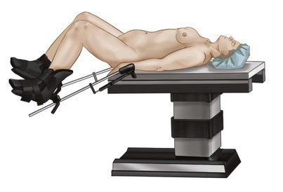
Patient positioning in the operating suite


Under general endotracheal anesthesia, the patient is positioned in lithotomy position with arms tucked at the sides to allow for surgeon mobility and to avoid brachial plexus injury (Fig. 37-2). A Foley catheter is placed. Two video monitors are placed at the foot of the bed. The cervix is grasped with a tenaculum and dilated with Hegar cones, and a uterine manipulator is inserted to assist with exposure and removal of the myomas. The surgeon stands on the patient’s left, the first assistant stands to the right, and the second assistant is between the legs. A Veress needle is inserted at the umbilicus, and the abdomen is insufflated with carbon dioxide to a pressure of 18 mm Hg.


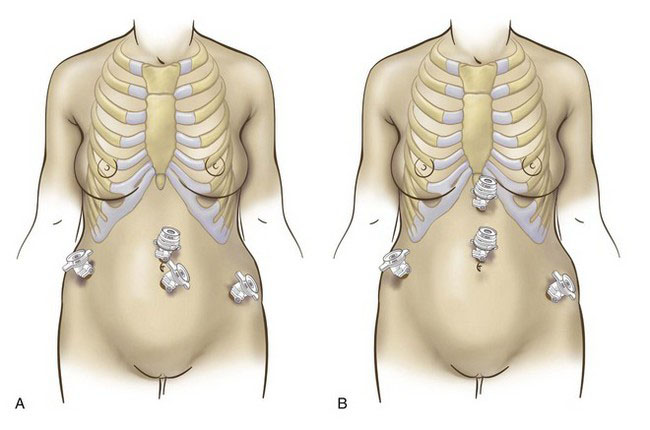
Positioning and placement of trocars


A 10-mm optical trocar is inserted at the umbilicus, and the laparoscope is placed through this port (Fig. 37-3A). Two 5-mm trocars are inserted in the lower abdomen, lateral to the inferior epigastric arteries. A third 5-mm trocar is inserted in the midline, level with or higher than the first two. If the patient has a large myoma that extends toward the umbilicus, then the surgeon may consider an open supraumbilical abdominal entry to establish pneumoperitoneum (Fig. 37-3B). The midline operative port then should be placed through the umbilicus or even higher. In the presence of large pathology, the optimal insertion points for the lateral 5-mm trocars can be determined after laparoscopic inspection of the pelvis.


< div class='tao-gold-member'>

Stay updated, free articles. Join our Telegram channel

Jul 20, 2016 | Posted by in GASTOINESTINAL SURGERY | Comments Off on Laparoscopic Myomectomy

Full access? Get Clinical Tree

Get Clinical Tree app for offline access