Laparoscopic Esophageal Mucosal Resection for High-Grade Dysplasia

Chapter 6 Laparoscopic Esophageal Mucosal Resection for High-Grade Dysplasia


The videos associated with this chapter are listed in the Video Contents and can be found on the accompanying DVDs and on Expertconsult.com.image


Barrett esophagus is defined as the metaplastic replacement of the normal squamous epithelium of the distal esophagus by columnar epithelium. Three histopathologic subtypes of metaplastic columnar epithelium have been described: two gastric phenotypes and one intestinal type. Because the intestinal type has the greatest risk for malignant transformation, the 2008 guidelines of the American College of Gastroenterology specify that the term Barrett esophagus should be restricted to columnar epithelium containing intestinal metaplasia. Estimates of the frequency of Barrett esophagus in the general population have ranged from 0.9% to 4.5%. Gastroesophageal reflux disease is the only known risk factor associated with the development of Barrett esophagus. A recent review of 15 epidemiologic studies in patients with gastroesophageal reflux disease (defined by at least weekly heartburn or acid regurgitation) identified a Barrett esophagus prevalence of 10% to 20% in the West and about 5% in Asia. Barrett esophagus with high-grade dysplasia is considered a premalignant condition. In patients with known Barrett esophagus, the annual risk for developing adenocarcinoma ranges from 0.2% to 2.0%.


The current gold standard for the treatment of Barrett esophagus with high-grade dysplasia is esophagectomy because of the perceived prevalence of invasive carcinoma in such specimens after esophagectomy. Recently, however, a meta-analysis of esophagectomy for high-grade dysplasia revealed invasive adenocarcinoma in only 12.7% of specimens. These data, along with inability or unwillingness to undergo esophagectomy, have further encouraged some patients to pursue more conservative treatment options for high-grade dysplasia. Other treatment options include endoscopic thermal therapy, photodynamic therapy, radiofrequency ablation, and laser ablation. Endoscopic mucosal resection has also been described as successful in treating high-grade dysplasia. One of the drawbacks of endoscopic mucosal resection for high-grade dysplasia or early esophageal cancer in Barrett esophagus has been the high rate of recurrent or metachronous lesions during follow-up in recent series (11% to 30%). Another drawback of endoscopic mucosal resection is that Barrett esophagus affecting segments longer than 2 cm is difficult to treat with endoscopic mucosal resection because piecemeal resection is often necessary. This usually requires a higher level of endoscopic expertise, multiple sessions, and an increased risk for complications. Additionally, it is difficult to be conclusive about the completeness of the resection at the lateral margins. This chapter presents a surgical alternative to esophagectomy and endoscopic management for high-grade dysplasia of the distal esophagus. The senior author (CTF) and colleagues previously published the success of laparoscopic transgastric esophageal mucosal resection; this chapter and the recording on the accompanying DVD (as well as on Expert Consult) are a follow-up to those published reports.


Positioning and placement of trocars


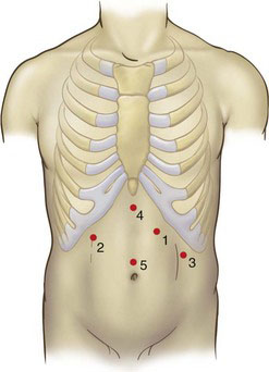
The patient is placed in a modified French lithotomy position with 30-degree reverse Trendelenburg (see Fig. 5-1 in Chapter 5). The surgeon stands between the patient’s legs, the camera operator stands to the patient’s right, and the first assistant stands to the patient’s left. Five trocars are placed as shown in Figure 6-1. The initial port (number 1) is placed using the optical trocar in the left midclavicular line subcostally and serves as the surgeon’s operating right hand. Subsequent trocars are placed in the order numbered two through five. The basic principles of trocar position and use should be followed: The camera port (number 5) is in the midline superior to the umbilicus. The surgeon’s left-hand working port (number 2) is placed in the right subcostal region in the midclavicular line. The retraction port for the assistant (number 3) is placed in the left midaxillary line caudad to port number 1. Trocar number 4 is used to retract the left lateral lobe of the liver and is placed in the subxiphoid region.


Operative technique


The left lobe of the liver is retracted with the use of an inflatable balloon retractor. The esophageal hiatus is visualized. If a hiatal hernia is present, the contents of the hernia are reduced by gentle traction, and the hernia sac is mobilized and excised. The esophagus is circumferentially mobilized and reduced into the abdomen for 3 to 5 cm. In addition, the esophagus is mobilized to an additional 5 cm in the mediastinum. This mobilization of the esophagus allows for a safer dissection of the esophageal mucosa later. If there is an inadvertent esophageal perforation, this may be diagnosed and addressed immediately. Following the circumferential mobilization of the esophagus, a 5-cm transverse gastrotomy is made 4 cm caudad to the gastroesophageal junction. The lumen of the esophagus and the location of the Z-line are visualized through the gastrotomy. A 30-degree laparoscope is indispensible in performing the operation.


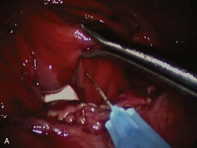
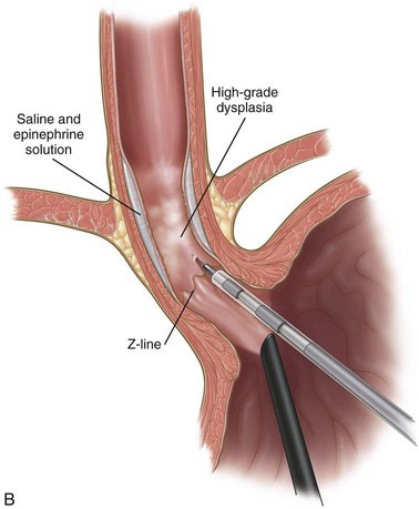
A solution of epinephrine and normal saline (1 : 100,000) is injected with a retractable hypodermic needle system at the Z-line of the distal esophagus to aid in the elevation of the mucosa (Fig. 6-2). The submucosal plane is entered with a modified hook electrocautery instrument; the hook is completely insulated except for the superior edge to allow contact with the underlying muscular layer and not cause thermal injury (Fig. 6-3). The mucosa is dissected further from the underlying smooth muscle with a curved laparoscopic spatula (Fig. 6-4). The mucosa is circumferentially dissected and excised in four quadrants. The mucosal segment is then excised using hook scissors. The tapered end of a lighted bougie (Medovations Inc., Milwaukee, Wis.) maneuvered by laparoscopic forceps is used as a retractor for exposing the four quadrants of mucosa (Fig. 6-5). The excised mucosa is oriented and marked for proximal and distal pathologic orientation. The raw surface of the esophagus is irrigated profusely, and any bleeding is controlled with cautious use of electrocautery. The esophageal wall in the area of mucosal resection is checked for intactness.


< div class='tao-gold-member'>

Stay updated, free articles. Join our Telegram channel

Jul 20, 2016 | Posted by in GASTOINESTINAL SURGERY | Comments Off on Laparoscopic Esophageal Mucosal Resection for High-Grade Dysplasia

Full access? Get Clinical Tree

Get Clinical Tree app for offline access