Laparoscopic Colectomy for Diverticulitis

Chapter 25 Laparoscopic Colectomy for Diverticulitis


The videos associated with this chapter are listed in the Video Contents and can be found on the accompanying DVDs and on Expertconsult.com.image


Colonic diverticular disease is common in Western society, occurring in about one third to two thirds of the population. It is hypothesized that a low-residue diet is a major causative factor. Age is another risk factor; diverticulosis is not uncommon in Americans in the fourth decade of life. Of patients with diverticulosis, as many as one fourth will develop diverticulitis.


The approach to the surgical management of diverticulitis depends greatly on the time of presentation. Many patients will present as a surgical emergency with the initial attack of diverticulitis. Historically, emergent surgery for diverticulitis was performed in either two or three stages. Since the 1980s, however, the practice of proximal diversion while leaving the diseased segment of colon behind (three-stage resection) has significantly decreased. Today, the Hartmann procedure is the most common two-stage operation. The initial stage of this operation is most commonly performed with the open approach; however, laparoscopic Hartmann procedure has been reported in the literature. More commonly, the second stage (i.e., Hartmann reversal) is performed laparoscopically. A single-stage approach to perforated diverticulitis has also been described. Additionally, with today’s potent antimicrobial regimens, the treatment of contained perforated diverticulitis with intravenous antibiotics only has been accepted as an option.


Operative indications


Elective colectomy for diverticulitis traditionally was reserved for the treatment of complications of the disease, such as recurrent attacks, perforation, abscess, fistula, stricture, or obstruction. Current recommendations are based on the clinical manifestations of the disease and the risk for recurrence and complications. It is advised that an elderly patient with a history of two episodes of diverticulitis or a younger patient (<50 years) with a history of one attack undergo an elective sigmoid colectomy to prevent a recurrent, more severe attack with the risks for complications. Both laparoscopic and open approaches for the elective treatment of diverticulitis have been described.


In this chapter, the technique for laparoscopic sigmoid colectomy with primary anastomosis in a complex case of diverticulitis is described. On the accompanying DVD (as well as on Expert Consult), we have included a video demonstrating the case of a patient with a history of multiple previous episodes of diverticulitis. The patient presented with a more severe recurrent attack, failed conservative treatment with percutaneous drainage of an abscess and intravenous antibiotics, and developed a colovesical fistula. The incidence of fistulas in patients with diverticular disease, the most common cause of colovesical fistula, is accepted to be 2%, although some centers have reported higher percentages. Colovesical fistulas are more common in males. Women who present with colovesical fistulas are commonly older or have a history of hysterectomy. The diagnosis is made clinically and confirmed with cystoscopy. This acute case can be compared with the case described in Chapter 15 of the Atlas of Minimally Invasive Surgery, 2009 (see Suggested Readings at the end of this chapter), in which the patient was successfully treated with antibiotics and the inflammation subsided before resection.


Preoperative evaluation


The preoperative evaluation of a patient undergoing a colectomy for diverticulitis is determined by the timing of the patient’s initial presentation.


When a patient presents with an acute attack of diverticulitis, a computed tomography (CT) scan to evaluate for perforation or abscess should be obtained. The patient is put on antibiotics covering aerobic and anaerobic bacteria. If an abscess is present, radiology-guided percutaneous drainage should be attempted. Patients with a microperforation may be managed conservatively with observation and antibiotics. If the inflammation improves with antibiotics, then the patient may undergo an elective resection.


The presence of a large perforation or diffuse peritonitis necessitates an urgent operation. Free colonic perforation secondary to diverticulitis is rare. Surgical management, whether open or laparoscopic, remains controversial.


If the patient presents with chronic diverticulitis, a barium enema to evaluate for stricture and fistula may be warranted in addition to a CT scan to evaluate for abscess and phlegmon.


For elective colectomy, a bowel preparation is recommended by most experts. The regimen consists of a traditional oral lavage with polyethylene glycol, followed by the administration of oral antibiotics (erythromycin and neomycin base) on the day before surgery. There is controversy surrounding the type of bowel preparation required and even whether bowel preparation is necessary. A single dose of a second-generation cephalosporin and metronidazole is given intravenously 30 minutes before incision. Sequential compression devices are placed for the prevention of deep venous thrombosis. Foley catheter placement for bladder decompression is required.


Patient positioning and port placement


For a laparoscopic sigmoidectomy, the patient is placed in the low lithotomy position with stirrups (Fig. 25-1). This positioning allows the end-to-end anastomotic (EEA) stapler to be used per the rectum. The left hip is elevated off the operating table by using a beanbag, viscoelastic cushion, or similar device. Placement of bilateral ureteral stents by a urologist, in particular infrared-illuminated stents (InfraVision, Stryker, San Jose, Calif.), is helpful in identifying the ureters when dealing with the phlegmon and inflammation of diverticulitis.


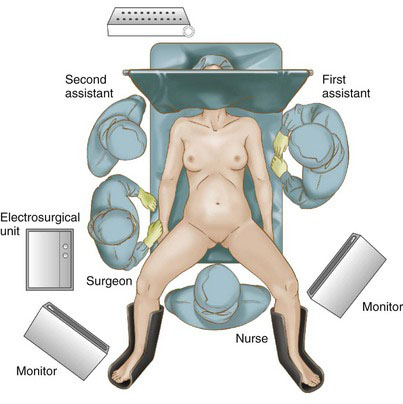
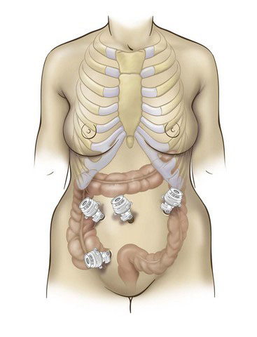
The surgeon stands on the patient’s right, facing a monitor. The authors prefer to access the abdomen using the open Hasson technique inferior to the umbilicus. This incision can then be extended to remove the specimen. If the patient has had previous surgeries, it may be prudent to insert a bladeless optical trocar at a different site to access the abdomen. After the pneumoperitoneum is established, two additional 12-mm trocars are placed in the right abdomen, just inferior and superior to the level of the umbilicus (Fig. 25-2). The use of 12-mm trocars at all port positions allows for maximal instrument flexibility. The placement of additional trocars may be necessary depending on the extent of the operation and the complexity of dissection. There are no set rules for the perfect port positioning. The surgeon must carefully consider each trocar placement in relation to the dissection and also to the other trocars. The main reason for conversion to open is the malpositioning of the trocar sites.


< div class='tao-gold-member'>

Stay updated, free articles. Join our Telegram channel

Jul 20, 2016 | Posted by in GASTOINESTINAL SURGERY | Comments Off on Laparoscopic Colectomy for Diverticulitis

Full access? Get Clinical Tree

Get Clinical Tree app for offline access