Laparoscopic Abdominoperineal Resection
KEY STEPS
1. Insertion of ports: 10-mm umbilical Hasson technique; 12-mm right iliac fossa; 5-mm left iliac fossa; 5-mm right upper quadrant.
2. Patient rotated to the right and placed in the Trendelenburg position.
3. Laparoscopic assessment and the small bowel and the omentum moved toward right upper quadrant.
4. Inferior mesenteric artery pedicle identified and dissected.
5. Left ureter identified and inferior mesenteric artery divided near origin.
6. Medial-to-lateral mobilization of the left colon.
7. Division of the left colon mesentery and left colon proximal to inferior mesenteric artery.
8. Rectal mobilization with total mesorectal excision.
9. Formation of trephine end colostomy in the left iliac fossa.
10. Wide perineal dissection, completion of rectal resection, and removal of specimen via perineum. Closure of perineal wound.
11. Closure of ports greater than 5 mm in size
ADDITIONAL ADVICE
1. Complete mobilization of the left colon is not required. Mobilization to allow formation of a left iliac fossa colostomy without tension is all that is necessary.
2. The left colon mesentery can be divided prior to freeing the lateral attachments of the sigmoid and left colon to aid retraction.
3. The levators are generally divided during the perineal dissection.
4. For patients with a tumor extending posteriorly, a prone approach is used to do the perineal dissection prior to the laparoscopic rectal mobilization.
5. In the perineal dissection, the tip of the coccyx is used as the posterior landmark and the pelvic cavity entered by dividing the levator ani muscle just anterior to it.
6. Special care must be taken during the anterior perineal dissection of the levator ani to protect the posterior surface of the vagina or prostate/urethra.
PATIENT POSITIONING
The patient is placed supine on the operating table on a beanbag. After induction of general anesthesia and insertion of an orogastric tube and a Foley catheter, the legs are placed in yellow fin stirrups (Fig. 21.1). Particular care is taken to ensure that the perineum is just below (distal) to the break in the table, so that good access can be obtained for the perineal dissection. The arms are tucked at the patient’s side and the bean bag is aspirated. The abdomen is prepared with antiseptic solution and draped routinely (see Chapter 2).
INSTRUMENT POSITIONING
The primary monitor is placed on the left side of the patient up toward the patient’s feet. The secondary monitor is placed on the right side of the patient at the same level and is primarily for the assistant during the early phase of the operation and port insertion (Fig. 21.2). The operating nurse’s instrument table is placed between the patient’s legs. There should be sufficient space to allow the operator to move from either side of the patient to between the patient’s legs if necessary. The primary operating surgeon stands on the right side of the patient with the assistant standing on the patient’s left, and moving to the right side, caudad to the surgeon once ports have been inserted. A 0-degree camera lens is used.
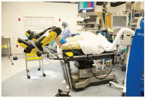 FIGURE 21.1. Patient position. |
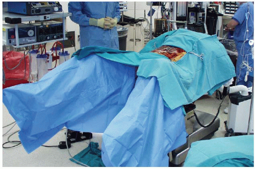 FIGURE 21.2. Room setup. |
UMBILICAL PORT INSERTION
This is performed using a modified Hasson approach (see Chapter 3). A vertical 1-cm subumbilical incision is made. This is deepened down to the linea alba, which is then grasped on each side of the midline using Kocher clamps. A scalpel (no. 15 blade) is used to open the fascia between the Kocher clamps and Kelly forceps are used to open the peritoneum bluntly. It is important to keep this opening small (<1 cm) to minimize air leaks. Having confirmed entry into the peritoneal cavity, a purse string of 0 polyglycolic acid is sutured around the subumbilical fascial defect (umbilical port site) and a Rommel tourniquet is applied. A 10-mm reusable port is inserted through this port site allowing the abdomen to be insufflated with CO2 to a pressure of 12 mmHg.
LAPAROSCOPY AND INSERTION OF REMAINING PORTS
The camera is inserted into the abdomen and an initial laparoscopy is performed carefully evaluating the liver, small bowel, and peritoneal surfaces. A 5-mm port is inserted in the right lower quadrant approximately 2 to 3 cm medial and superior to the anterior superior iliac spine. This is carefully inserted lateral to the inferior epigastric vessels, paying attention to keep the tract of the port going as perpendicular as possible through the abdominal wall. A 5-mm port is then inserted in the right upper quadrant at least a hand’s breadth superior to the lower quadrant port. A 12-mm left lower quadrant port is also inserted at the colostomy site, through which the endoscopic stapler can be passed to divide the intestine. Again all of these remaining ports
are kept lateral to the epigastric vessels. This may be insured by diligence to anatomical port site selection and using the laparoscope to transilluminate the abdominal wall prior to making the port site incision to identify any obvious superficial vessels.
are kept lateral to the epigastric vessels. This may be insured by diligence to anatomical port site selection and using the laparoscope to transilluminate the abdominal wall prior to making the port site incision to identify any obvious superficial vessels.
DEFINITIVE LAPAROSCOPIC SETUP
The assistant now moves to the patient’s right side, standing caudad to the surgeon. The patient is rotated with the left side up and right side down, to approximately 15 to 20 degrees tilt, and often as far as the table can go. This helps to move the small bowel over to the right side of the abdomen. The patient is then placed in the Trendelenburg position. This again helps gravitational migration of the small bowel away from the operative field. The surgeon then inserts two atraumatic bowel clamps through the two right-sided abdominal ports. The greater omentum is reflected over the transverse colon so that it comes to lie on the stomach. If there is no space in the upper part of the abdomen, one must confirm that the orogastric tube is adequately decompressing the stomach of gas. The small bowel is moved to the patient’s right side allowing visualization of the medial aspect of the rectosigmoid mesentery. This may necessitate the use of the assistant’s 5-mm atraumatic bowel clamp through the left lower quadrant in order to tent the sigmoid mesentery cephalad.
Related posts:


Stay updated, free articles. Join our Telegram channel

Full access? Get Clinical Tree


