, Mark Thomas1 and David Milford2
(1)
Department of Renal Medicine, Birmingham Heartlands Hospital, Birmingham, UK
(2)
Birmingham Children’s Hospital, Birmingham, UK
Abstract
In this chapter we explain:
The basic anatomy and physiology of the kidney
How kidney function changes through life
The Anatomy of the Kidney
The kidneys are complex and beautiful organs. Their internal structure is revealed by anatomical studies using light and electron microscopy (Figs. 1.1, 1.2, 1.3, 1.4 and 1.5).
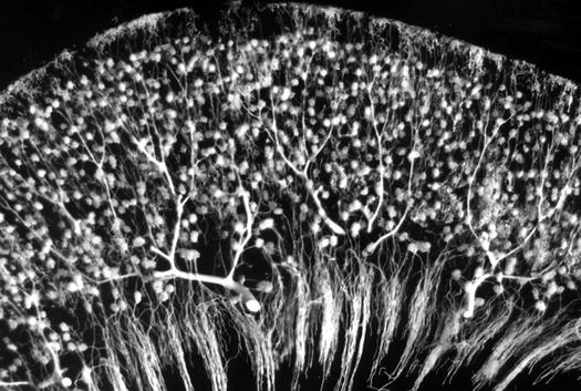
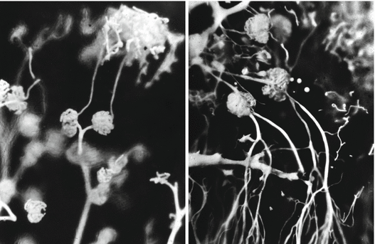
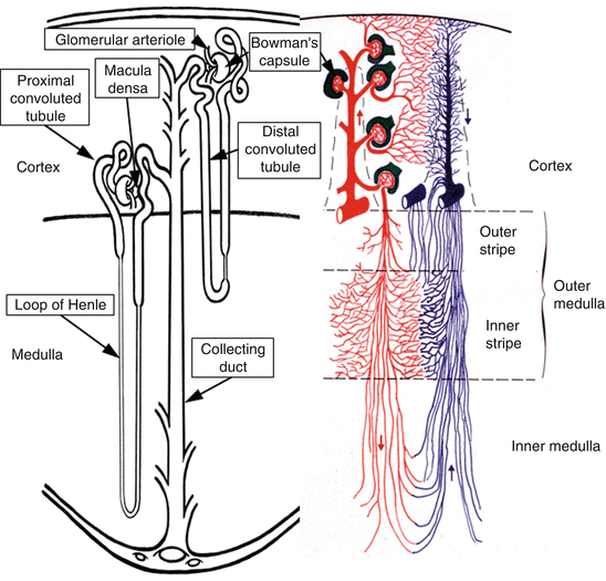
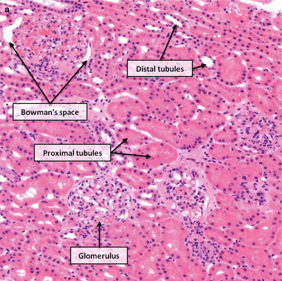
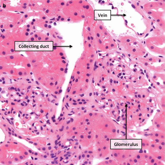
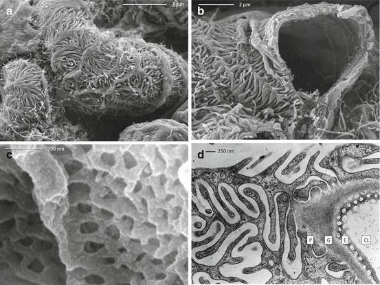
Fig. 1.1
Longitudinal section through the cortex and outer medulla of a rabbit kidney in which the artery has been injected with white Microfil. Microfil has filled the arteries, arterioles, glomerular tufts and the early part of the post-glomerular capillaries in the cortex and outer medulla (Courtesy of Dr Lise Bankir, Centre de Recherche des Cordeliers, Paris, France)
Fig. 1.2
Rabbit kidney injected with white Microfil through the renal artery. Left: detail of a longitudinal section showing a small part of the superficial cortex. The glomerular tufts of two superficial glomeruli are visible with their post-glomerular capillaries located in the very superficial cortex. Right: detail of a longitudinal section showing part of the deep cortex and the outer stripe of the outer medulla. The glomerular tufts of two juxtamedullary glomeruli are visible with their efferent arterioles that run towards the outer medulla where they give rise to vascular bundles (Courtesy of Dr Lise Bankir, Centre de Recherche des Cordeliers, Paris, France)
Fig. 1.3
Nephons and their blood supply. Left: a short looped- and a long looped-nephron. Right: the different vascular territories and their location in the four renal zones. For clarity, the cortex has been widened and the inner medulla compressed
Figs. 1.4
(a, b) Light micrographs of normal renal cortex with the main structures indicated. Haematoxylin and eosin; a ×100, b ×200
Fig. 1.5
Scanning electron micrographs of a mouse glomerular capillary. (a) The surface of a capillary showing podocyte (5000× by SecretDisc). (b) A cut open capillary revealing the endothelial lining (10,000× by SecretDisc). (c) The inner surface showing fenestrations in the endothelial cells (100,000× by SecretDisc). (d) Transmission electron micrograph of a section of glomerular capillary wall showing the layers that form the glomerular filtration barrier. CL capillary lumen, E endothelial cell fenestrations, G glomerular basement membrane, P podocyte slit diaphragm (Image d made available by James D. Jamieson and the Department of Cell Biology, Yale University School of Medicine. Original 3.25 in. × 4 in. lantern slides were scanned at 600 dpi. Original magnification ×16,000. The original work has been cropped and modified with labels) [1]
Turning Blood into Urine
The kidneys are central to homeostasis [2]. Through exquisite sensory mechanisms [3] they regulate blood pressure, water [4], sodium [5], potassium [6], acidity [7], bone minerals [8], and haemoglobin. But their core function is the excretion of the waste products of metabolism in urine.
About 22 % of cardiac output goes to the kidneys and about 20 % of the plasma is filtered, producing about 170 L of glomerular filtrate per day. Ninety-nine percent of this is reabsorbed as it flows along the nephrons so only about 1.5 L of urine is produced per day.
Filtration occurs through the glomerular filtration barrier [9] . This is made up of five layers [10] (Fig. 1.6):
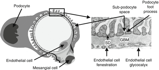
Fig. 1.6
The cells and five structural components that form the glomerular filtration barrier. SD slit diaphragm, GBM glomerular basement membrane
the glycocalyx covering the surface of the endothelial cells
holes (fenestrations) in the glomerular endothelial cells
the glomerular basement membrane
the slit diaphragm between the foot-processes of the podocytes
the sub-podocyte space between the slit diaphragm and the podocyte cell body
The composition of glomerular filtrate is determined by the structure, arrangement and electrical charge of the collagen protein molecules that form the filtration barrier. So glomerular filtration is both size-selective and charge-selective; molecules that are too large or too highly charged cannot get through.
A substantial amount of albumin does get through the barrier, between 3.3 and 5.7 g per day. A proportion passes from the sub-podocyte space through the podocytes by transcytosis [11]. Passage of albumin through the barrier is increased by angiotensin II. Almost all the filtered albumin is reabsorbed by active uptake into the proximal tubular cells [12].
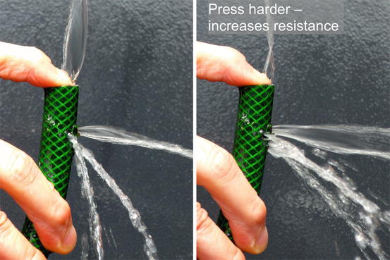
A simple model of the haemodynamics of glomerular filtration can be made from a garden hose (Fig. 1.7).