Author
Reason for exclusion
Elbahnasy [2]
N = 13
Menezes [3]
N = 22
Dasgupta [4]
SFR for stone size not reported and unclear follow-up protocol
Breda [5]
N = 15
Chung [6]
N = 12
Ferrandino [7]
Not peer reviewed
Lahme [8]
Not peer reviewed
Riley [29]
N = 22
Sejiny [9]
Stone series within calyceal diverticula
Desai [10]
N = 18
Yili [11]
N = 13
A total of 18 studies with 1,362 patients met our inclusion criteria, including 566 patients in early series (Table 8.2) and 796 in contemporary series (Table 8.3). Of these, 90 patients were the result of “prospective” series with SFRs based on CT scan follow-up (see Table 8.3). Both of these (prospective randomized controlled trial Pearle et al. [12] and prospective trial Portis et al. [13]) are reviewed in greater detail in discussion section of this chapter. The remaining 16 studies were retrospective in nature and report a wide range of non-standardized follow-up protocols. It is important to note a significant lack of uniformity in outcome reporting among all trials. Therefore, SFRs from these studies were quite variable and have a high potential for bias and unreliability. Seven studies used more than one imaging modality to calculate SFR without stratifying patients per that modality, consequently making results unclear [14–20]. CT was the imaging modality of choice in 4/9 “modern” series (see Table 8.3), while kidneys–ureters–bladder films (KUB) the most common radiograph in the early series (7/9 studies, Table 8.2). Detection of residual stone burdens solely by CT scan was performed by three groups [12, 13, 21], while CT was combined with a variety of imaging modalities in two studies [15, 18]. Five studies have been published with a focus primarily on ureteroscopic treatment of lower pole stones (Tables 8.2 and 8.3) [12, 15, 22–24]. Four of these studies were published prior to widespread use of ureteral access sheaths in 2003, and two investigated patients who had previously failed SWL and/or had residual stones following PCNL [17, 25].
Table 8.2
Results from early ureteroscopy series
Author | N | Mean size (cm) | Imaging modality | Median F/U | SFR | Notes |
|---|---|---|---|---|---|---|
Fabrizio [20] | 100 | 0.81 | KUB, US, IVP | 1 and 3 mos | 77% | SFR includes 56 patients with concurrent ureteric calculi and includes fragments <3 mm |
Grasso [19] | 45 | >2.0 | KUB | 3 mos | 81% | 45 stones (not patients) treated at 3 medical centers; SFR not stratified by center or by patient |
Tawfiek [14] | 59 | 0.68 | KUB, US | Post-op to 3 mos | 80% | Some patients had post-op KUB only as f/u data |
El-Anany [16] | 30 | >2.0 | KUB, US | 1 mo | 77% | Mean stone size NR; SFR includes residual calculi of <2 mm; 17/19 patients were stone free >6 mos |
Sofer [30] | 56 | 1.35 | KUB | 1–2 wks | 84% | 56/598 had renal stones only; unclear how SFR was calculated |
Grasso [22] | 90 | NR | US | 3 mos | 82% (<1 cm) 71% (1–2 cm) 65% (>2 cm) | 90 lower pole stones (not patients) treated at 3 medical centers; 70/90 stones had f/u data available |
Kourambas [15] | 34 | NR | CT, IVP | 3 mos | 85% | 30/34 patients had LP stones < 1.5 cm |
Hollenbeck [23] | 60 | 0.87 | KUB | <1 mo | 79% | Unclear number of ureteric stones and total follow-up; 21% of patients had stones >1 cm |
Schuster [24] | 78 | 0.86 | KUB | 1 mo | 76% | 16/72 pts had concomitant ureteral stones |
Table 8.3
Results from contemporary ureteroscopy series
Author | N | Mean size (cm) | Imaging modality | Median F/U | SFR | Notes |
|---|---|---|---|---|---|---|
Stav [25] | 81 | 0.92 | KUB and US | 3 wks | 46% | 24/81 patients with ureteric and renal stones; all patients failed previous SWL |
Pearle [12] | 32 | <1.0 | CT | 3 mos | 50% | Randomized prospective trial comparing SWL and URS |
Holland [17] | 93 | 0.91 | KUB and US | 3 mos | 73% | 11/93 patients with ureteric and renal stones; 43 patients failed previous SWL; 8 patients failed previous PCNL |
Johnson [31] | 143 | NR | US | 3 mos | 90% (<1.0 cm) 89 % (1–2 cm) 75% (>2 cm) | n = 10 for stones <1 cm, n = 100 for stones 1–2 cm, n = 33 for stones >2 cm |
Portis [13] | 33 | 0.94 | CT | 1 mo | 54% | Prospective trial; 25/58 patients with ureteric and renal stones; SFR based solely on renal stones |
Coccuza [21] | 44 | 1.15 | CT | 2 mos | 93% | 15/44 patients had ureteric and renal stones; SFR based solely on renal stones |
Alcaide [32] | 100 | 1.5 | IVP | 3 mos | 93% | 95/128 stones treated were lower pole; 96% of patients had 3 mos follow-up |
Hyams [18] | 120 | 2.4 | CT, KUB and US | 2 mos | 47% | Results from 3 different surgeons at 3 different centers; SFR not stratified by center nor imaging modality |
Herrera-Gonzalez [27] | 125 | 1.2 | URS | Post-op and 1 day | 74% | SFR calculated from direct visualization at case end; stone size ranged 2 mm–5 cm |
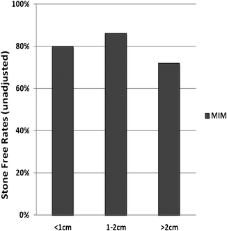
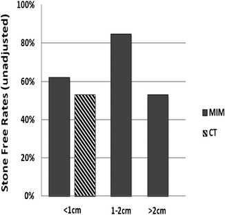
Using data from Tables 8.2 and 8.3, we created early and contemporary URS, Figs. 8.1 and 8.2. The number of patients was taken from each tabled study and multiplied by their reported SFR. These were summed and divided by total number of patients from all included studies, yielding an average. For example, five studies were reported before 2003 with a mean stone size <1 cm [14, 20, 22–24]. Total patient number multiplied by SFR and divided by total patient number resulted in the following: [(47 × 0.94) + (100 × 0.77) + (59 × 0.80) + (78 × 0.76) + (60 × 0.79)/(47 + 100 + 59 + 78 + 60)], equaling 79.9% which rounded to 80% (Fig. 8.1). All reported SFRs are termed “unadjusted” as they are pooled samples from a variety of studies and imaging modalities. Any use of imaging other than strict CT criteria (i.e., studies that combined CT and a variety of other imaging modalities) was termed “multiple imaging modality” or MIM.


Fig. 8.1
Ureteroscopy stone-free rates, early studies. Average weighted stone-free rates reported by retrospective case series published prior to 2003 using multiple imaging modalities (MIM), including KUB, IVP, US, CT. Stone size <1 cm = 80%, 1–2 cm = 86%, and >2 cm = 72%. SFR includes all studies regardless of patient number in series

Fig. 8.2
Ureteroscopy stone-free rates, contemporary studies. Average weighted stone-free rates reported from both retrospective and prospective studies published during or after 2003 using either multiple imaging modalities (MIM—including KUB, IVP, US, CT) or strict CT criteria. Stone size <1 cm = 63% (MIM) and 53% (CT), 1–2 cm = 85%, and >2 cm = 53%. SFR includes all studies regardless of patient number in series
Outcomes in Ureteroscopy: SFR, Size, Location, and Default
Stone-Free Rate: CT Criteria
Since the advent of minimally invasive surgery, the definition of “success” in the management of kidney stones has undergone a series of transformations. Until the mid-1980s, open or percutaneous surgical series considered any residual stone an operative failure. With the widespread use of SWL in the late 1980s, small “clinically insignificant fragments” became an accepted term, as long as the residual stones were less than 4 or 5 mm in size, asymptomatic, and noninfected. Even today, there remains no consensus within the urologic community as to what constitutes a “clinically insignificant” fragment. Therefore, most urologists agree that attempts should be made to render a patient completely stone-free at the time of the first procedure, as any residual stone fragment may require continued radiological monitoring and may have the potential for future growth or symptoms.
Stay updated, free articles. Join our Telegram channel

Full access? Get Clinical Tree


