Fig. 13.1
Electrohydraulic lithotrite
Due in part to the higher risk of ureteral perforation, the use of EHL for the treatment of ureteral stones was largely supplanted by the other modalities discussed within this section. EHL (as well as PL and USL) was further marginalized as the equipment for SWL and LL became more economically competitive and the techniques demonstrated high success rates and more favorable safety profiles [17, 18].
Pneumatic Lithotripsy
PL was introduced in the early 1990s with the development of the Swiss Lithoclast (Microvasive-Boston Scientific Corp., Boston, Massachusetts). This simplified means of stone fragmentation is accomplished via a rigid energy probe that is placed in direct contact with the stone (Fig. 13.2). The application of compressed air to a sliding metal projectile in the hand piece transmits energy to the probe and subsequently to the surface of the stone, facilitating stone fragmentation in a manner similar to that of a jackhammer [19]. Depending upon probe diameter and the air pressure setting, the rigid probe of the Lithoclast achieves a maximum velocity of 12.1–33.3 m/s with a maximum probe displacement of 2.53 mm [20]. The use of a readily available energy source (compressed air) as well as reusable probes and hand pieces made PL a practical and economical treatment option for ureteral stones upon its introduction [21].
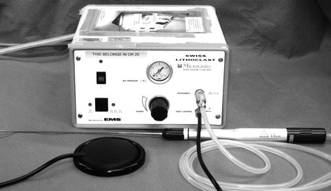
Fig. 13.2
Pneumatic lithotrite
The early rigid PL probes of the Lithoclast had to be used with a rigid ureteroscope or nephroscope, which initially restricted the application of the technique. To address this limitation, flexible pneumatic probes were developed for the Lithoclast. Although these new probes could be used through flexible ureteroscopes, a progressive loss of probe tip displacement, velocity, and energy, as well as stone fragmentation efficacy resulted, particularly with deflection angles greater than 24° [22, 23]. A rival PL system, the Browne Pneumatic Impactor (BPI; Browne Medical Systems, Minneapolis, MN), was the first lithotrite to come to market with flexible probes for PL. Utilizing the same basic principal as the Lithoclast, the BPI used compressed air to propel a 1.5–1.8 F nickel–titanium (Nitinol) wire through a polymer sheath that could be employed through a semirigid or actively deflectable flexible ureteroscope. Normal probe tip displacement and stone fragmentation efficacy was only maintained through 30 ° of deflection with the smallest flexible BPI probes, but larger flexible probes maintained tip displacement and velocity and were able to fragment even hard stones, at 90 ° of deflection [24]. In a small series of patients with stones as large as 20 mm, 7 of 8 ureteral stones were successfully fragmented using the BPI [25].
In addition to the maneuverability limitations imposed by PL probes, proximal stone fragment migration was a pervasive problem inherent to the nature of PL stone fragmentation. As many as 18% of patients undergoing PL for treatment of ureteral stones required secondary procedures due to proximal migration of large stone fragments [6, 26]. In an attempt to overcome this problem, as well as to provide an efficient means of small stone fragment evacuation, the Lithovac (Microvasive-Boston Scientific, Boston, Massachusetts) was developed. The Lithovac is a hollow suction probe through which the rigid Lithoclast energy probe projects. The device provides constant suction and is designed to facilitate the simultaneous evacuation of smaller fragments as the stone is destroyed, as well as helping to prevent proximal migration of larger fragments that would necessitate a secondary procedure. In a study performed shortly after its introduction, the Lithovac achieved a 100% ureteral stone fragmentation rate, with no proximal fragment migration and a stone-free rate of 95% [27].
Ultrasonic Lithotripsy
The first practical application of ultrasound took place in France in 1916 when Chilowsky and Langevin patented the use of ultrasonic sonar for the underwater localization of submarines [30]. Ultrasonic energy is produced when alternating electrical current is applied to plates on the opposing sides of a crystal. The resultant mechanical vibrations of the crystal propagate waves of energy at a constant frequency, as determined by the voltage applied to the plates and the nature of the crystal. Observations of the disruptive effects of the first sonar on marine life led to the elucidation of the mechanism by which the ultrasonic vibrations affect objects within their path. The high frequency energy waves of ultrasound alternately compress and pull on particles within the radiation field. In transmission media such as water, this results in cavitation. When these cavities rapidly collapse instantaneous pressure is produced with magnitude and force capable of destroying solid objects [31]. Early attempts to fragment urinary and biliary stones by the direct application of ultrasound were unsuccessful due to the high electrical energy requirements and difficulties in conducting and focusing the ultrasonic energy [32–34]. In the early 1970s the ultrasonic lithotrite was developed. Rather than relying on the direct application of ultrasound energy and the resultant cavitation to disintegrate stones, the ultrasonic lithotrite uses an ultrasound transducer to rapidly vibrate a hollow probe and longitudinally transmit mechanical energy to the surface of stone, causing fragmentation in the same manner a drill (Fig. 13.3) [31]. The hollow USL probe facilitates simultaneous suction removal of stone fragments and constant-flow irrigation aids in fragment removal and cools the USL probe to reduce the risk of thermal damage to adjacent tissues [35].
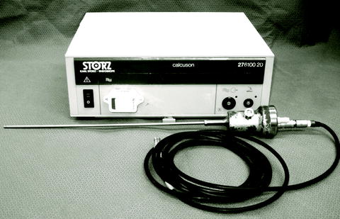
Fig. 13.3
Ultrasonic lithotrite
Prior to its adoption for ureteroscopy, USL was widely employed in the transurethral treatment of bladder stones and in the percutaneous treatment of renal stones [36, 37]. Initially, USL in the ureter was performed in a blind fashion with fluoroscopic guidance. Results were more favorable than those seen with blind treatment of ureteral stones with EHL or PL, and complications rates were much lower [4]. Direct vision ureteroscopic USL was made possible by the miniaturization of ureteroscopes that could employ the first generation 4.5–6 F hollow probes. The development of a 2.5 F solid wire USL probe significantly increased the treatment range and efficiency of the technique [35]. The wire USL probe transmits the mechanical energy of the ultrasonic transducer to the stone in a transverse nature which, unlike the longitudinal energy transmission of a hollow probe, resulting in disintegration of the stone. A multi-institutional series of 118 upper, mid, and lower ureteral stones treated with USL demonstrated an overall success rate of 96.6% with a low rate of complications and secondary procedures [38].
As in the case of EHL and PL, SWL and LL systems have largely replaced USL in the treatment of ureteral stones, but they remain useful in some clinical situations such as patients with steinstrasse or a large ureteral stone burden [39].
Combination Ultrasonic and Pneumatic Lithotripsy
A combination intracorporeal lithotripter, the Lithoclast Ultra (Boston Scientific Corporation, Natick, MA) was introduced to take advantage of the rapid stone fragmentation of pneumatic lithotripsy and the efficient fragment evacuation ability of a hollow ultrasonic lithotripter. The pneumatic ultrasonic lithotripsy (PUL) allows the dual energy sources to be used individually or concomitantly depending on operator preference, stone composition, size, or location. Laboratory tests on stone phantoms found more efficient stone fragmentation and evacuation with the use of the combined energies at their highest settings, when compared to the highest setting operation of either mode alone [40, 41]. Early clinical experience verified the utility of PUL, but a randomized comparison revealed stone-free rates achieved with PUL were not significantly greater than those of USL alone [42].
The Safety of Intracorporeal Lithotrites
The emergence of each new lithotripsy technology prompted studies evaluating and comparing the risks associated with the use of their novel energy sources within the narrow confines of the ureter. The introduction of SWL and LL further highlighted the risks associated with all preceding methods of intracorporeal lithotripsy, and renewed concerns about the acute and long-term risks of their use. We provide a brief survey of the results of in vitro and in vivo analyses of the safety of each technique.
Electrohydraulic Lithotripsy
Among the methods of intracorporeal lithotripsy discussed in this chapter, EHL has the highest risk of acute tissue damage when used to treat ureteral stones. Although considered safe and effective for the treatment of bladder stones, early studies on the ureters of cadavers demonstrated the Urat-1 was capable of rapidly inflicting tissue damage and ureteral perforation [11]. Direct application of the Urat-1 to the abdominal wall and bladder mucosa of anesthetized canines was found to cause blistering and hemorrhage through the full thickness of the dermis and epidermis of the abdomen, and similar effects through the mucosa and detrussor muscle of the bladder [12]. High speed video of spark discharges at the tip of EHL probes revealed that the blast energy of the cavitation bubbles cause only small areas of trauma, limited in size to the diameter of the probe, even with the probe in direct contact with sections of opened human ureter. However, when confined within in the lumen of the ureter, the blast energy cannot be effectively dispersed and it causes rapid expansion of the ureteral wall and complete disruption of the ureter in a longitudinal fashion, as far as 1 cm from the tip of the probe [43]. To better characterize the immediate and potential long-term ureteral damage of EHL, the probe of a second generation EHL lithotrite was directly applied in a perpendicular fashion to the ureters of two anesthetized pigs. Histological samples were then analyzed at the time of injury, 1 day, and 6 days after injury. Within the ureter EHL produced an immediate partial abrasion of the urothelium and caused edema within the underlying lamina propria. By day one the urothelium was totally abraded and the edema in the lamina propria was increased and extended into the ureteric smooth muscle. Analysis of the ureteral sections 6 days after injury revealed worsening damage and reactive hyperplasia with excessive mucous secretion. This extensive damage can be partially attributed to the initial blast injury caused by the rapid expansion and collapse of the plasma bubble. To an even greater degree, full-thickness ureteral damage is due to the heat that is rapidly generated at the probe tip [44].
In an ex vivo model utilizing sections of pig ureter, application of EHL at the lowest power setting produced visible perforation of the ureter within approximately 25 pulses [45]. To reduce the risk of collateral tissue damage, a plasma shield was developed for the EHL probe. Composed of a stainless steel coil and a perforated end cap, the plasma shield was designed to fit the distal end of the EHL probe and confine the plasma within the coil. The energy was then focused through a hole in the end of the cap that reduced energy transfer to the tissue. In a study employing rabbit bladders, an unshielded EHL probe perforated the bladder in two pulses, compared to a mean of 35 pulses required to perforate with a shielded EHL probe [46].
While the EHL remains an efficient and economical tool for the treatment of some bladder stones, the propensity for tissue damage has limited its role for treatment of stones within the ureter.
Pneumatic Lithotripsy
In contrast to the early outcomes of EHL, PL the novel Swiss Lithoclast appeared to be effective and safe for use in the treatment of ureteral stones. Deliberate attempts to perforate the ureters of anesthetized female swine with 0.8 and 1.0 mm rigid Lithoclast probes resulted in perforation with the 0.8 mm probe, but not until after delivery of 250 consecutive shocks. Perforation of the ureter with the 1.0 mm probe was deemed impossible. Although no macroscopic lesions were visible on the ureteral epithelium, abrasion, and edema were seen on histologic analysis of treated sections of ureter [44]. Similar areas of transmural edema, abrasion, and hemorrhage were identified microscopically in sections of pig ureter subjected to direct firing of a 3.5 F Lithoclast probe for 5–7 s. In the same experiment, animals sacrificed 3 weeks later had no evidence of ureteral perforation on retrograde pyelography, and treated areas of ureteral tissue showed no microscopic evidence of damage. Animals sacrificed at 6 weeks posttreatment also had normal retrograde pyelograms and no direct evidence of microscopic tissue trauma to treated ureteral sections [47]. The safety of PL was further confirmed when the direct application of a 3 F rigid probe for 6 min of continuous activation failed to perforate ex vivo sections of pig ureter, even with “vigorous attempts to impale the ureter” [45].
An extensive study of the safety of PL, including its use to treat plaster-of-Paris stone phantoms in swine ureters and stones in the kidneys, ureters, and bladders of human patients, revealed that even direct and deliberate application to soft tissues results in minimal trauma. Among the 37 ureteral stones treated in the human portion of the study, one patient suffered a minor ureteral perforation attributed to “overenthusiastic ureteroscopy.” Follow-up intravenous urograms of 25 patients obtained 1 month after PL demonstrated no ureteral injuries [48].
Ultrasonic Lithotripsy
Some of the earliest experiments to assess the effects of USL on the tissue of the ureter were performed in dogs. A 30-s application of the USL probe to human stones implanted in the canine ureter was followed by direct application of the probe to the ureteral mucosa for 5, 10, and 15-s intervals. Gross and histological examination of the ureteral tissue was performed after 48 and 72 h. No gross trauma to the ureteral wall was noted during applications of 10 s or less. Applications of the USL probe directly to the ureteral wall for 15 s caused visible and histologic changes consistent with thermal damage. The investigators theorized that this thermal damage could easily be prevented during routine use of the USL when pulses of 10 s or less are used and the buildup heat at the probe tip is avoided. Because this study was conducted prior to the introduction of ureteroscopy and direct vision intracorporeal lithotripsy, the main cause of most ureteral perforation was actually excessive force applied to the probe, and not ultrasound energy or the resultant heat [34]. A second study examining the effect of USL on the canine bladders revealed that direct contact application of the probe to the urothelium for 10 min produced only modest long-term histologic changes in sections examined 1 month after treatment [31]. A test of the ability of USL probe to cause perforation of swine ureter after 12 pulses of 3 s each resulted in no perforations and no macroscopic changes in the urothelium. Histologic examination of treated tissue on the day of treatment revealed no changes to the urothelium. Microscopic changes seen 1 and 6 days after treatment were moderate and less extensive than those seen in tissue samples of ureters treated with PL, EHL, or LL. The authors concluded that USL was among the safest of the lithotripsy technologies available at the time [44].
Because the energy of USL is produced by the generation of sound waves, its clinical use presented a unique safety concern regarding the effects of USL on the hearing of patients, physicians, and operating room personnel. In the US workplace noise exposure limits are established and monitored by the Occupational Safety and Health Administration (OSHA). The allowable continuous exposure is 8 h at 90 decibels (dBA), with a 50% decrease in duration mandated for each 5 dBA above 90 dBA. Measurements of the sound levels of the three commercially available USL units during actual cases of intracorporeal lithotripsy showed both decibel levels and exposure durations well below OSHA standards. Additionally, an audiogram performed on a urologist with a 3-year total of more than 40 cases of ultrasonic lithotripsy did not detect any hearing threshold sensitivity deficit [49]. An experimental model used to simulate the use of USL in the ureter during pregnancy concluded that the peak pressure of sound emitted during lithotripsy was unlikely to harm the hearing of the fetus [50].
Comparative Studies of Outcomes, Complications, and Cost
In addition to concerns regarding the safety of emerging lithotrite technologies, the introduction of each successive modality instigated comparisons to existing modes with special interest in the outcomes, complications, and costs. In this section we present summaries of a representative selection of these studies. Although LL is not discussed in the chapter, studies comparing various types of lasers to EHL, PL, USL, and PUL are also included because the laser would eventually become the dominate energy source for intracorporeal ureteral lithotripsy. Table 13.1 summarizes the comparative studies detailed below.
Table 13.1
Summary of comparative studies
Year author | n | Lithotrites (n) | Outcome measures and results |
|---|---|---|---|
1991 Vicente J | 100 | USL (67) EHL (33) | •Successful fragmentation (USL vs. EHL): 93 vs. 73% •Three patients each group required secondary procedure •1 EHL patient required ureteral reimplant |
1994 Naqvi S | 200 | PL (152) LL (48) | •Non-fragmentation rate (PL vs. LL): 1 vs. 10% •Stone free ≤4 weeks (PL vs. LL) : 95 vs. 84% |
1995 Hofbauer J | 72 | EHL (34) PL (38) | •Successful fragmentation (EHL vs. PL): 85 vs. 90% (p = 0.12) •Need for secondary SWL (EHL vs. PL): 15 vs. 11% (p = 0.63) •Ureteral perforation (EHL vs. PL): 18 vs. 3% (p < 0.01) |
1997 Teichman J | 70 | EHL (23) LL (47) | •Stone free at procedure (EHL vs. LL): 56 vs. 94% (p = 0.002) •Stone free at 3 months (EHL vs. LL): 87 vs. 98% (p = 0.01) |
2001 Sun Y | 286 | PL (145) LL (140)
Related posts: Informed Consent and Perioperative Antibiotics
Complex Anatomy: Horseshoe, Pelvic, and Malrotated Kidneys Informed Consent and Perioperative Antibiotics
Complex Anatomy: Horseshoe, Pelvic, and Malrotated Kidneys
 Flexible Ureteroscopy: Holmium:YAG Laser and Optical Fibers Flexible Ureteroscopy: Holmium:YAG Laser and Optical Fibers
 Radiation Safety During Ureteroscopy Radiation Safety During Ureteroscopy
 Semirigid Ureteroscopy Step-by-Step: The Tulane Approach Semirigid Ureteroscopy Step-by-Step: The Tulane Approach
 Upper Tract Urothelial Carcinoma: Ureteroscopic Biopsy and Specimen Preparation Upper Tract Urothelial Carcinoma: Ureteroscopic Biopsy and Specimen Preparation
Stay updated, free articles. Join our Telegram channel
Full access? Get Clinical Tree
 Get Clinical Tree app for offline access

|